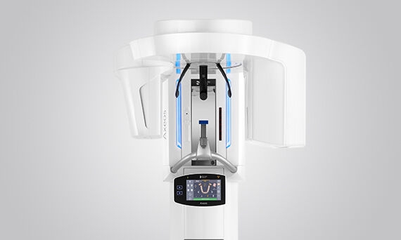
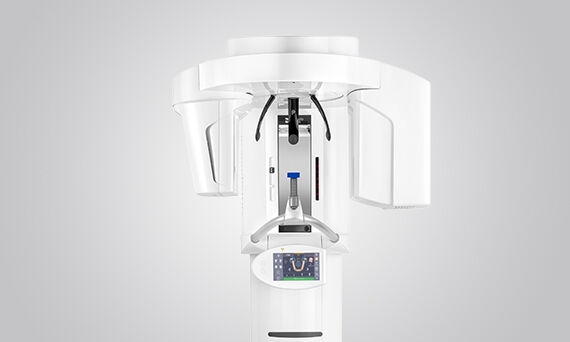
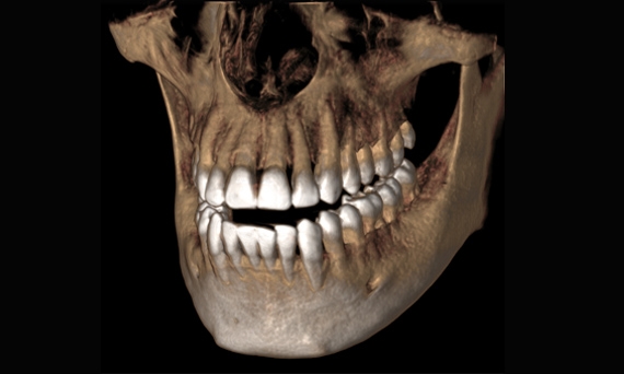
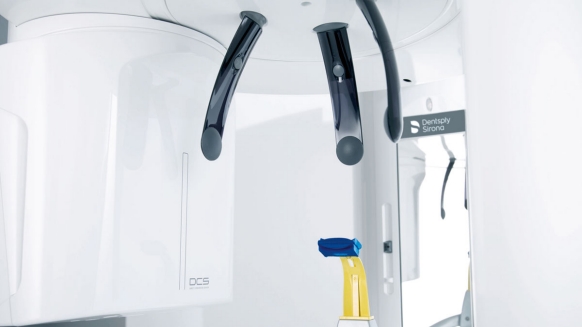
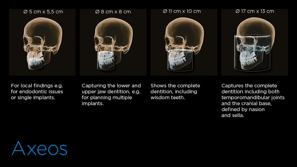
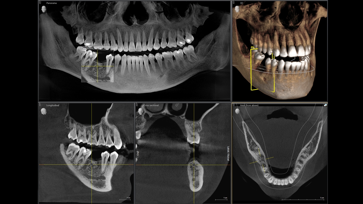
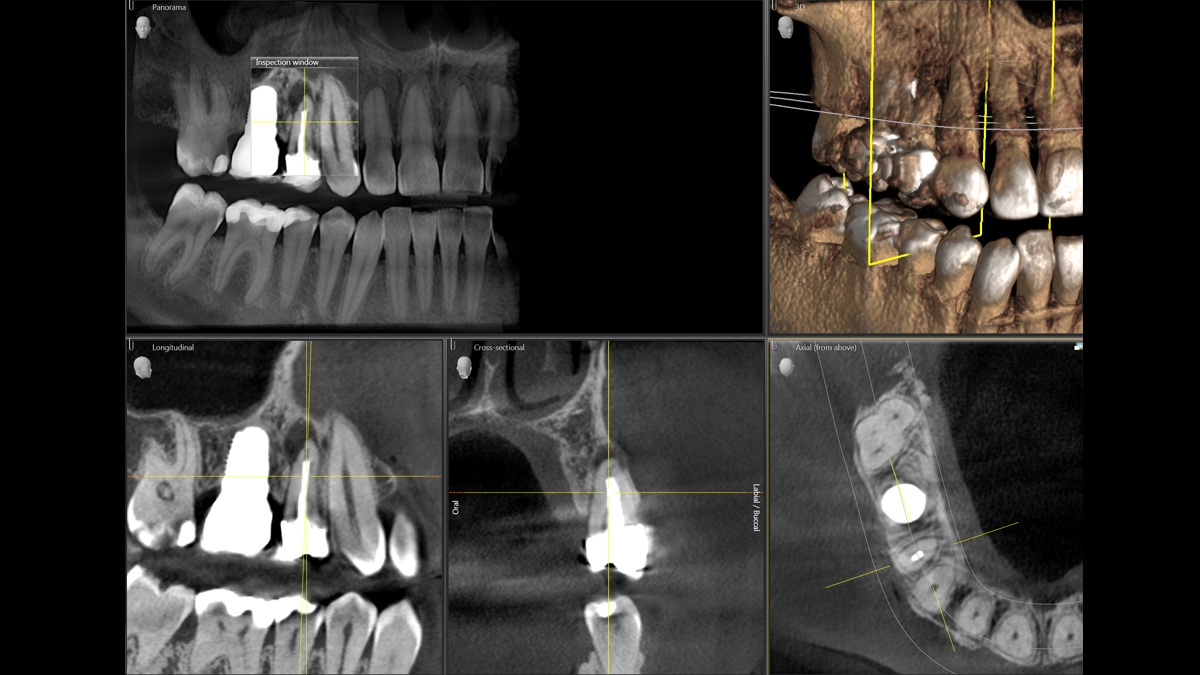
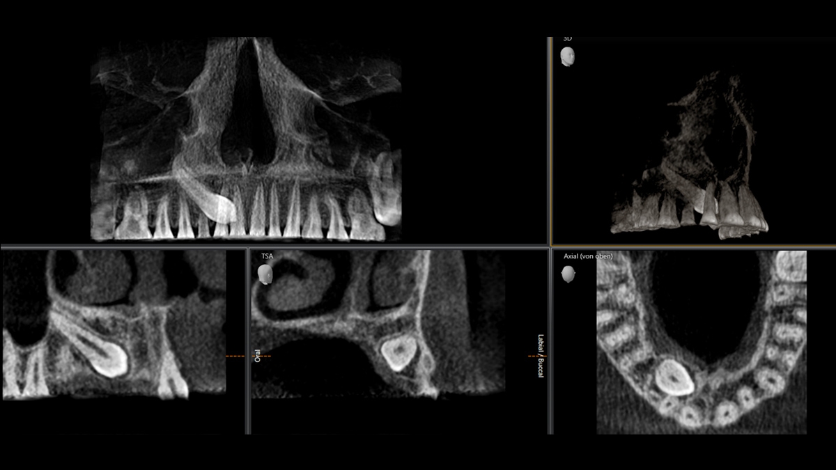
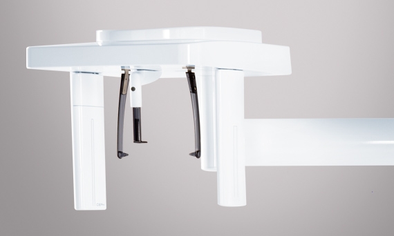
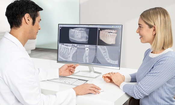
3D Imaging

More precise diagnoses, clearer explanations: 3D imaging has many benefits. Be it overlaid teeth, unexpected nerve channel paths, concealed roots, or temporomandibular joint anomalies, the value 3D images provide in diagnostics is unmeasurable.

image-comparison grip-lines arrow-top-right zip arrow-right video minus arrow-back text shop twitter file home quick-order arrow-left youtube search arrow-down check all filter site_references image Slice 1 zoom 🎬 icons / _ icon size / _ download-queue Created with Sketch. chevron-down arrow-right picture pdf podcast exe plus hotline close xxx-1 burger clipboard linked-in ppt Artboard 🎬 icons / _ icon size / _ download-queue_add Created with Sketch. Layer 1 Created by potrace 1.15, written by Peter Selinger 2001-2017 My Account press_releases Created with Sketch. PRESS ds-instruments ds-preventive ds-implants ds-endodontics Cart presentation Created with Sketch. DS-Preventive-Icon-Grey video Created with Sketch. other Created with Sketch. text_piece Created with Sketch. promotion Created with Sketch. Spare Parts ds-imaging-systems case_study Created with Sketch. ds-treatment-centers broschure Created with Sketch. ds-prosthetics ds-restorative video Created with Sketch. flyer Created with Sketch. whitepaper Created with Sketch. article Created with Sketch. image Created with Sketch. Service ds-cad-cam Artboard Quick Order Dentist Chair study Created with Sketch. ds-orthodontics ds-implants ds-endodontics ds-cerec digital-scan grinder ds-diagnosis END-Website-Icons-Shaping-Cleaning-Obturation step_1 analyse ds-abutment-placement more-quality initiative scan more-quality ds-polishing digital ds-irrigation ds-treatment-centers Test END-Website-Icons-Shaping-Cleaning-Obturation treatment ds-restorative ds-implant-planning realistic construct book ds-planning-impression END-Website-Icons-Shaping-Cleaning-Obturation END-Website-Icons-Shaping-Cleaning-Obturation mirror anwender ds-preventive planning-2 ds-obturation ds-cad-cam 04 ds-implant-placement ds-retention-protection ds-anesthetics restauration form ds-inlab oven CORP-icon-grey-screen-tooth-cavity-pulpa robot plan place ds-orthodontics PDF-Formular_ganzneu PDF overlay ds-bone-regeneration ds-treatment-preparation vorher_nachher ds-maging-systems wifi END-Website-Icons-Shaping-Cleaning-Obturation omnicam PDF-Formular_neu_vonGünther treatment consulting planning comparison ds-set-up ds-prosthetics support step_3 ds-tooth-preparation ds-treatments ds-scaling step_2 ds-instruments software quote-english quote-french quote-text-german Created with Sketch. upload Created with Sketch. version Created with Sketch. globe EndoEquipmentGreypng Imaging Grey 2svg EndoGlideShapingGreypng irrigation blue Imaging 1svg Imaging 2svg DS-Preventive-Icon-Grey Final_Implants_Category_Implant-Systems Access Blue obturation blue restoration restoration grey Access Grey treatment EndoEquipmentBluepng irrigation grey Diagnosis primeprint-grey Hub Bluesvg Final_Implants_Procedure_Single-tooth Diagnsis blue Imaging blue EndoRetretmentBluepng restoration blue Final_Implants_Procedure_Edentulous-therapy Final_Implants_Procedure_Hard-and-soft-tissue-management obtruation grey endo grey treatment blue Imaging blue 1svg Imaging grey EndoGlideShapingBluepng EndoRetretmentGreypng polishing Hubsvg Final_Implants_Category_Implant-Dentistry polishing blue Diagnosis Grey Cerec Bluesvg Imaging Blue 2svg treatment grey Hub Greysvg Scaling Cerec Greysvg endo blue polishing grey search Imaging Grey 1svg scaling blue Cerecsvg DS Points Logo Imaging scaling grey

Custom 3D Imaging

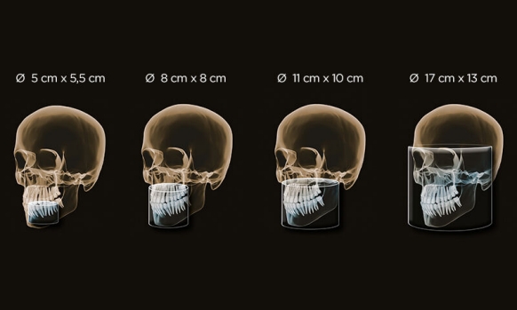
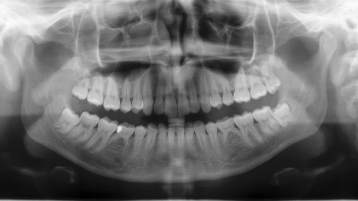
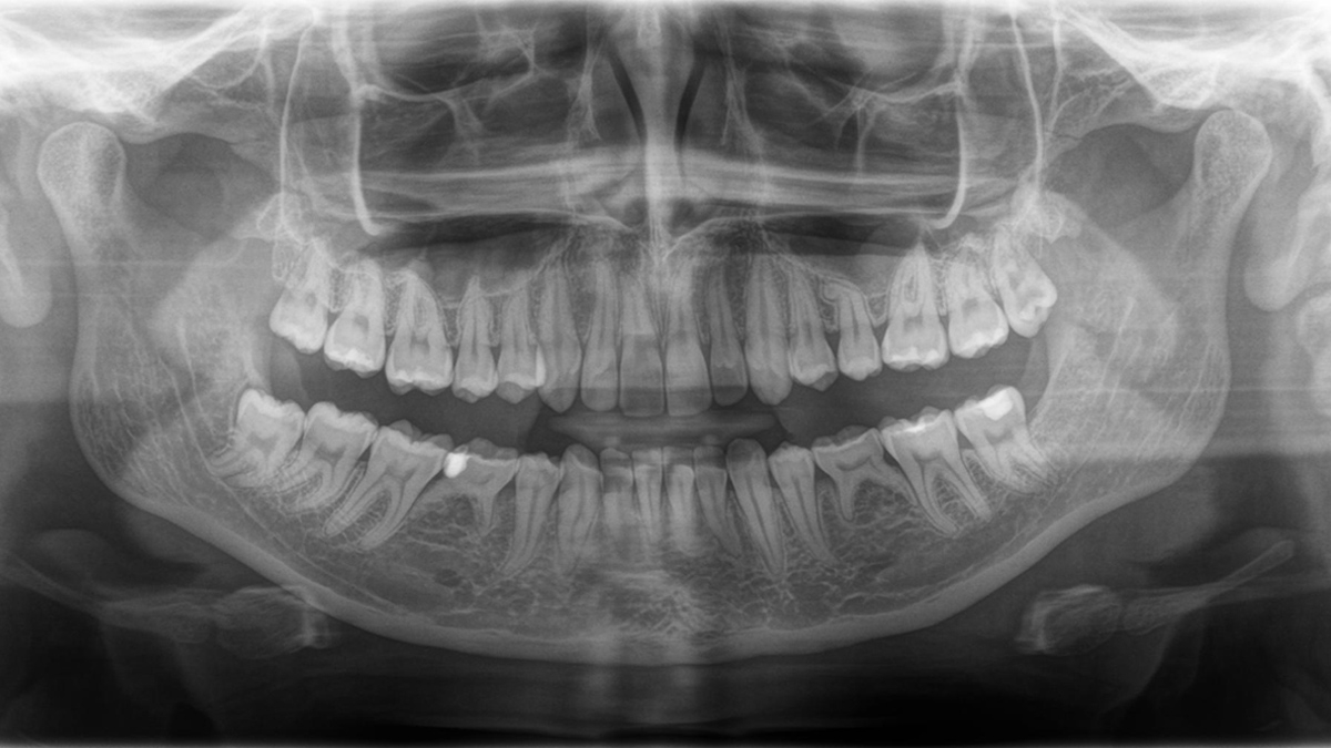
Examine a precise area with a FoV of Ø 5 cm x 5.5 cm or evaluate the complete dentition including temporomandibular joints with Ø 17 cm x 13 cm. Whether HD images in high resolution or low-dose images in the dose range of 2D images, our systems adapt to your individual patient case. These are just some of the advantages you receive with our 3D X-ray solutions.


3D Units

Axeos

The versatile 2D/3D hybrid unit with a big FOV and high image quality for practices with a wide treatment spectrum. Axeos convinces not only with its high-calibre performance, but also with its focus on comfort and design as well.

Orthophos S 3D

This All-round 2D/3D hybrid unit offers a full range of capabilities to support all of the 2D and 3D tasks in your practice. Thanks to its flexible volume sizes and adjustable dose options, you have a great partner to support your workflows.

Benefits at a Glance

Axeos and Orthophos are based on a proven successful hardware platform and software system. This gives you:

Volumes according to your diagnostic needs

From a focused field of view for endodontic questions (Ø 5 cm x Ø 5.5 cm) up to a large field of view (Ø 17 cm x Ø 13 cm) that covers complete dentition, including both TMJs and the cranial base, defined by nasion and sella.

Efficient diagnostics thanks to exceptional image quality

Always following the ALARA principle, our 2D/3D hybrid solutions make sure that you can count on the image quality you need for the given indication at the lowest possible dose.

Broader practice offering with modern treatment solutions

Our smart treatment solutions are perfect companions for our x-ray units, giving you new possibilities for your practice offering, now and into the future.

Keep your practice running reliably thanks to proven quality

Proudly "Made in Germany" quality and more than 100,000 units sold worldwide, we can say one thing for sure: Our Extraoral Imaging Solutions stand for best-in-class reliability.

Reproducible results thanks to easy handling

Thanks to our Patient Positioning & Image Assistant concept, easy positioning and X-ray capture are simple.


Feature Highlights of our 2D/3D Hybrid Family:

Variety of volumes

Our fields of view have been developed to match your needs. Starting with a focused 5 cm x 5.5 cm FoV for endodontic or single implant cases up to 17 cm x 13 cm for the complete dentition and upper airway analysis, you decide which volume is the right one for you.

No shine on MARS

Metal artifacts present a challenge for 3D imaging. With three-dimensional reconstruction, radiopaque objects create shadows and streaking effects on the X-ray image. This is especially true for patients with metal restorations, these artifacts make diagnostics more problematic. The MARS software (metal artifact reduction software) reduces metal artifacts in 3D images significantly. This improves the image quality substantially and renders diagnostics with less complication.  

High definition mode (HD)

At standard settings, Dentsply Sirona imaging units are set to SD (Standard definition) mode with a supreme balance of image quality and dose value for everyday practice life. For certain indications, i.e. visualisation of fine root canals, high definition (HD) mode shows all necessary details at just the click of a button.

Patient Positioning & Image Assistant concept

We developed a 10-point concept for easy patient positioning and X-ray imaging. Our concept is primarily about two things: high image quality and comfort for the patient and the assistant. This concept supports and provides

3D Imaging: Experience the Difference

3D technology is becoming increasingly well-established in dental practices around the world. For more precise diagnoses and treatment explanations, 3D X-rays offer a number of advantages. Whether with superimposed teeth, unexpected nerve canal paths, hidden roots, or temporomandibular joint anomalies, 3D images are invaluable for a large number of diagnoses.

With the Axeos and Orthophos S from Dentsply Sirona, several hundred individual images are produced when taking a 3D X-ray in the space of just a few seconds. Using this data, the software generates a 3D image of the jaw area. The practitioner is then able to take a "virtual tour" of the scanned volume and assess the data from every dimension.

2D Panoramic Imaging: Revolutionary Technologies

Even though 3D X-rays are becoming increasingly established in a growing number of dental practices, in many cases 2D imaging still has a rightful place. This is largely due to radiation hygiene, which is a core component of modern dental diagnostics. In terms of improvements in image quality, the options available in 2D technology are far from exhausted. 2D X-ray devices from Dentsply Sirona score highly in daily handling, thanks to technical innovations for unparalleled sharp images and operation that leaves nothing to be desired.

Panoramic image quality is constantly evolving

To the left: 10 year old PAN image. To the right: DCS image taken with Orthophos SL


Cephalometric Imaging

Orthophos XG 3D and Orthophos S 3D both offer the capability to install a cephalometric arm at any time. To be sure it fits in your X-ray room, the arm can be mounted either on the right side or left side of the unit. 

Ceph programs that cover all orthodontic needs

Using a dedicated sensor, you get lateral and symmetrical p.a. or a.p. as well as carpus images. In cases of displaced teeth, you can also fall back on the benefits of 3D X-rays to determine their exact location.

Technical Data

Overview Chart

Performance features
Axeos Orthophos S 3D
Fields of View (cm) 5x5 to 17x13 5x5 to 8x8
5x5 to 11x10
3D Low Dose yes yes
HD mode yes yes
Smart height adjustment  yes -
X-ray generator
60 - 90 kV, 3-16mA 60 - 90 kV, 3-16mA
Panoramic exposure time
P1: max 14,2 s
P1 Quickshot: max 9,1 s 
P1: max 14,2 s
P1 Quickshot: max 9,1 s
Radiation time Ceph
Standard 9,4 s
Quickshot 4,7 s
Standard 9,4 s
Quickshot 4,7 s
User interface
new EasyPad EasyPad
Patient positioning
automatic (occlusal bite block) automatic (occlusal bite block) 
Panorama technology
DCS CsI Plus
Autofocus
yes yes
Ceph arm (optional)
left or right left or right
Ceph unit with 2 sensors
yes yes
Quickshot
yes yes
Base
optional optional
Wheelchair accessible
yes yes
Remote control
optional optional
Ambient Light yes (cabinet and backlight) -

FAQ

The requirements actually follow those for the image processing software, Sidexis 4.

Yes, the Axeos works exclusively with Sidexis 4. But data migration from Sidexis XG to Sidexis 4 is very easy. 

Axeos units feature a characteristic, compact design to fit even in small x-ray rooms with ease.


  Orthophos XG 3D Axeos
Minimum space need
(depth x width x height)
1.411 x 1.280 x 2.250 mm 1.411 x 1.280 x 2.250 mm

Downloads

The 3D X-ray Family 4 MB

Find out more about our 3D and 2D/3D Hybrid product range and download our 3D family brochure here.

Orthophos SL - Cases in practice 3 MB

Want to find out what your colleagues are doing already with the different Orthophos SL features and how the unit is opening up their practice for new offerings? Download our user case brochure here.


Contact us for more information

Find out more about the imaging systems from Dentsply Sirona and request information on intraoral imaging, software or 2D and 3D imaging technology.