Orthophos S 3D

Creating the optimal workflow – this hybrid X-ray unit ensures just that with a full range of capabilities for all the 2D and 3D tasks in your practice and thanks to the flexible volume sizes and dose options.

image-comparison grip-lines arrow-top-right zip arrow-right video minus arrow-back text shop twitter file home quick-order arrow-left youtube search arrow-down check all filter site_references image Slice 1 zoom 🎬 icons / _ icon size / _ download-queue Created with Sketch. chevron-down arrow-right picture pdf podcast exe plus hotline close xxx-1 burger clipboard linked-in ppt Artboard 🎬 icons / _ icon size / _ download-queue_add Created with Sketch. Layer 1 Created by potrace 1.15, written by Peter Selinger 2001-2017 My Account press_releases Created with Sketch. PRESS ds-instruments ds-preventive ds-implants ds-endodontics Cart presentation Created with Sketch. DS-Preventive-Icon-Grey video Created with Sketch. other Created with Sketch. text_piece Created with Sketch. promotion Created with Sketch. Spare Parts ds-imaging-systems case_study Created with Sketch. ds-treatment-centers broschure Created with Sketch. ds-prosthetics ds-restorative video Created with Sketch. flyer Created with Sketch. whitepaper Created with Sketch. article Created with Sketch. image Created with Sketch. Service ds-cad-cam Artboard Quick Order Dentist Chair study Created with Sketch. ds-orthodontics ds-implants ds-endodontics ds-cerec digital-scan grinder ds-diagnosis END-Website-Icons-Shaping-Cleaning-Obturation step_1 analyse ds-abutment-placement more-quality initiative scan more-quality ds-polishing digital ds-irrigation ds-treatment-centers Test END-Website-Icons-Shaping-Cleaning-Obturation treatment ds-restorative ds-implant-planning realistic construct book ds-planning-impression END-Website-Icons-Shaping-Cleaning-Obturation END-Website-Icons-Shaping-Cleaning-Obturation mirror anwender ds-preventive planning-2 ds-obturation ds-cad-cam 04 ds-implant-placement ds-retention-protection ds-anesthetics restauration form ds-inlab oven CORP-icon-grey-screen-tooth-cavity-pulpa robot plan place ds-orthodontics PDF-Formular_ganzneu PDF overlay ds-bone-regeneration ds-treatment-preparation vorher_nachher ds-maging-systems wifi END-Website-Icons-Shaping-Cleaning-Obturation omnicam PDF-Formular_neu_vonGünther treatment consulting planning comparison ds-set-up ds-prosthetics support step_3 ds-tooth-preparation ds-treatments ds-scaling step_2 ds-instruments software quote-english quote-french quote-text-german Created with Sketch. upload Created with Sketch. version Created with Sketch. globe EndoEquipmentGreypng Imaging Grey 2svg EndoGlideShapingGreypng irrigation blue Imaging 1svg Imaging 2svg DS-Preventive-Icon-Grey Final_Implants_Category_Implant-Systems Access Blue obturation blue restoration restoration grey Access Grey treatment EndoEquipmentBluepng irrigation grey Diagnosis primeprint-grey Hub Bluesvg Final_Implants_Procedure_Single-tooth Diagnsis blue Imaging blue EndoRetretmentBluepng restoration blue Final_Implants_Procedure_Edentulous-therapy Final_Implants_Procedure_Hard-and-soft-tissue-management obtruation grey endo grey treatment blue Imaging blue 1svg Imaging grey EndoGlideShapingBluepng EndoRetretmentGreypng polishing Hubsvg Final_Implants_Category_Implant-Dentistry polishing blue Diagnosis Grey Cerec Bluesvg Imaging Blue 2svg treatment grey Hub Greysvg Scaling Cerec Greysvg endo blue polishing grey search Imaging Grey 1svg scaling blue Cerecsvg DS Points Logo Imaging scaling grey

A True All-rounder of its Class

The high-quality 2D/3D x-ray device with a comprehensive range of services for every practice. Whether as a pure 2D device or including a 3D module – the Orthophos S is a reliable partner and optimised for everyday tasks. Its CsI sensor with autofocus function ensures clear images, even in anatomically difficult cases. The patented occlusal bite block automatically positions patients. For use in orthodontics, the Orthophos S is also available with an optional ceph arm. And because future-proofing is important to Dentsply Sirona, the cephalometric arm can be retrofitted at any time.


Your Advantages at a Glance

With the Orthophos S 3D, your practice is equipped for a broad range of clinical tasks in 2D and 3D.

Sharp and autofocused images

Even in anatomically difficult cases with the 2D CsI sensor with autofocus function

Selectable volume sizes

From ø 5 cm x 5.5 cm to ø 11 cm x 10 cm

3D according to your needs

With the Low Dose and HD function, you can produce 3D images in the dose range of 2D X-ray and HD images with up to 80 μm resolution.

Ceph arm on the left or right

For ceph images, can be ordered as an option or can be retrofitted at any time

Maximum consistency and reproducibility

Thanks to automatic patient positioning with the patented occlusal bite block

Safe and proven patient positioning

With motorised temple and forehead support, automatic temple width measurement, light localisers and sturdy handles

Orthophos S 3D – Highlights


Easy to Use, Safe Positioning

For you, using an x-ray devise is about two things: The best possible image and having your patient feel comfortable. For both, the Orthophos S 3D offers unique, patented solutions: 

  • The intuitive user interface of the EasyPad
  • Automatic positioning aids such as the occlusal bite block and the 3-point head fixation
  • An integrated temple width measurement

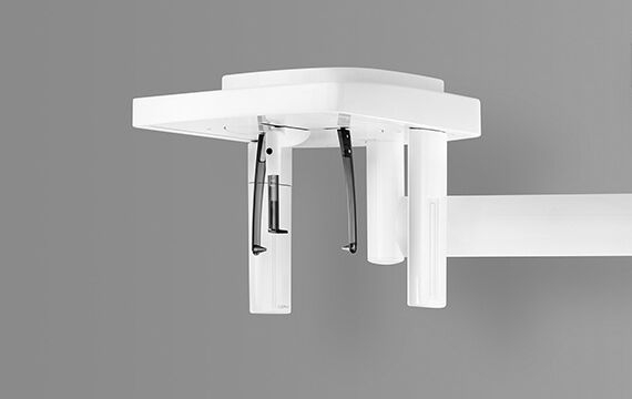
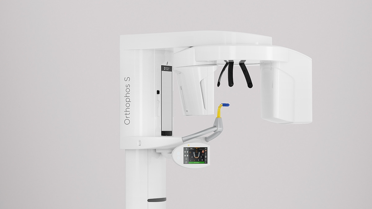
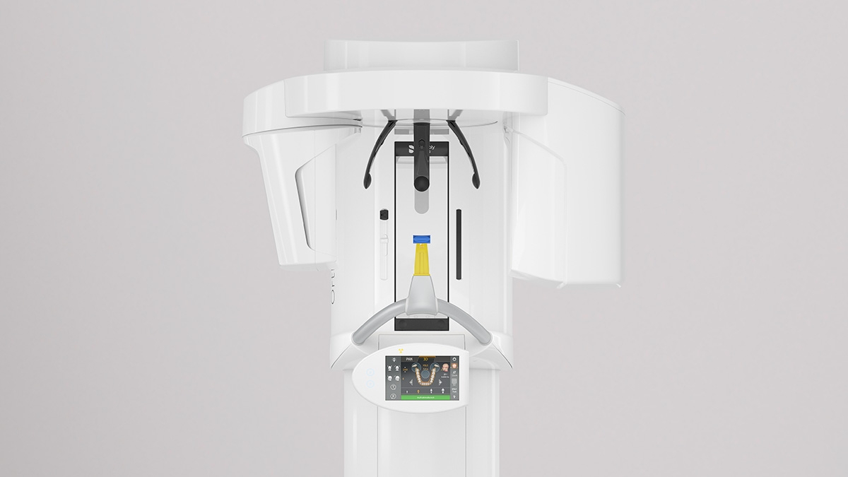
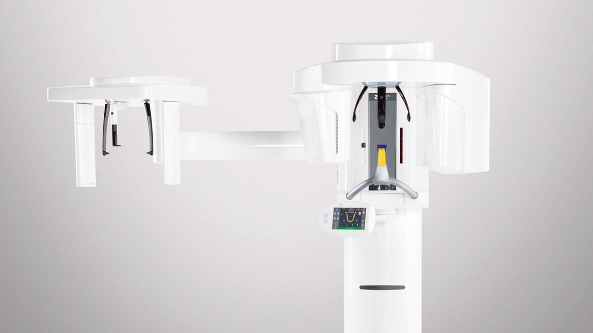
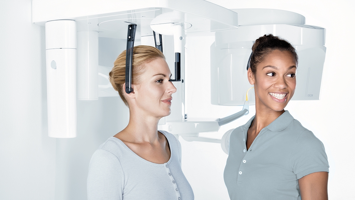
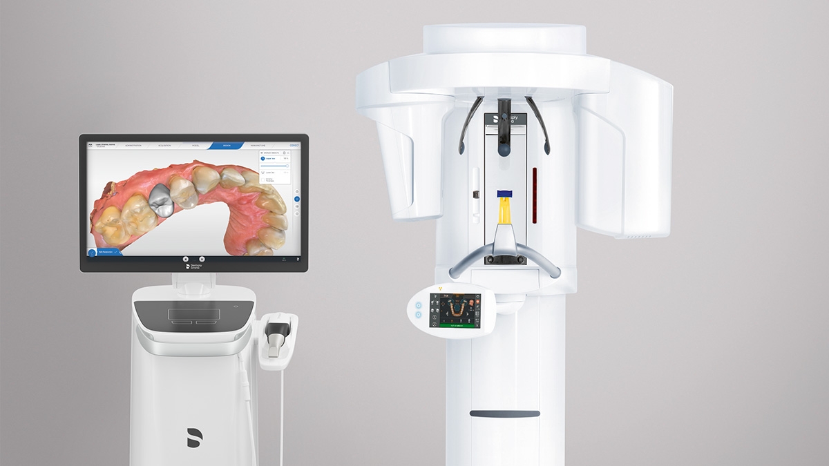
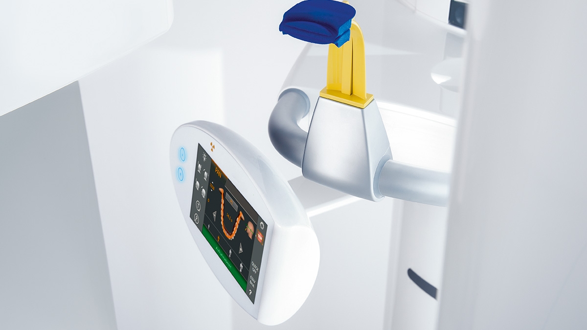
Patient Positioning & Image Assistant concept

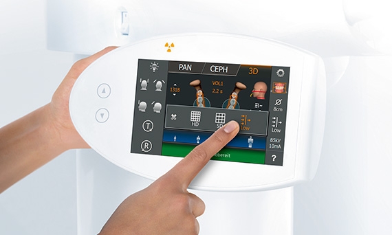
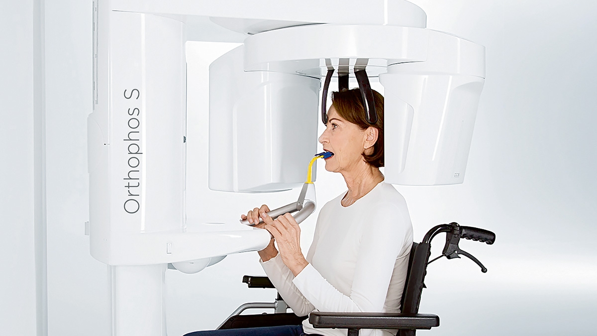
We developed a 10-point concept for easy patient positioning and X-ray imaging. Our concept is primarily about two things: high image quality and comfort for the patient and the assistant. This concept supports and provides


Cephalometric Imaging

Orthophos S 3D offers the capability to install a cephalometric arm at any time. And to be sure it fits in your x-ray room, the arm can be mounted either on the right side or left side of the unit.

Ceph programs that cover all orthodontic needs

Using a dedicated sensor, you get lateral and symmetrical p.a. or a.p. as well as carpus images. For displaced teeth, you can also use 3D x-rays to determine their exact location.


FAQ

The requirements actually follow those for the image processing software, Sidexis 4.

Yes, the Axeos works exclusively with Sidexis 4. But data migration from Sidexis XG to Sidexis 4 is very easy. 

Axeos units feature a characteristic, compact design to fit even in small x-ray rooms with ease.


  Orthophos XG 3D Axeos
Minimum space need
(depth x width x height)
1.411 x 1.280 x 2.250 mm 1.411 x 1.280 x 2.250 mm

The Extraoral X-Ray Family

Find out more about our 2D and 2D/3D hybrid product range and download the extraoral X-ray family brochure here.

Dentist with Axeos unit in the background

Technical Data

Overview Chart

Performance features
Axeos Orthophos S 3D
Fields of View (cm) 5x5 to 17x13 5x5 to 8x8
5x5 to 11x10
3D Low Dose yes yes
HD mode yes yes
Smart height adjustment  yes -
X-ray generator
60 - 90 kV, 3-16mA 60 - 90 kV, 3-16mA
Panoramic exposure time
P1: max 14,2 s
P1 Quickshot: max 9,1 s 
P1: max 14,2 s
P1 Quickshot: max 9,1 s
Radiation time Ceph
Standard 9,4 s
Quickshot 4,7 s
Standard 9,4 s
Quickshot 4,7 s
User interface
new EasyPad EasyPad
Patient positioning
automatic (occlusal bite block) automatic (occlusal bite block) 
Panorama technology
DCS CsI Plus
Autofocus
yes yes
Ceph arm (optional)
left or right left or right
Ceph unit with 2 sensors
yes yes
Quickshot
yes yes
Base
optional optional
Wheelchair accessible
yes yes
Remote control
optional optional
Ambient Light yes (cabinet and backlight) -

Contact us for more information

Find out more about the imaging systems from Dentsply Sirona and request information on intraoral imaging, software or 2D and 3D imaging technology.