inEos X5 Scan with inLab

image-comparison grip-lines arrow-top-right zip arrow-right video minus arrow-back text shop twitter file home quick-order arrow-left youtube search arrow-down check all filter site_references image Slice 1 zoom 🎬 icons / _ icon size / _ download-queue Created with Sketch. chevron-down arrow-right picture pdf podcast exe plus hotline close xxx-1 burger clipboard linked-in ppt Artboard 🎬 icons / _ icon size / _ download-queue_add Created with Sketch. Layer 1 Created by potrace 1.15, written by Peter Selinger 2001-2017 My Account press_releases Created with Sketch. PRESS ds-instruments ds-preventive ds-implants ds-endodontics Cart presentation Created with Sketch. DS-Preventive-Icon-Grey video Created with Sketch. other Created with Sketch. text_piece Created with Sketch. promotion Created with Sketch. Spare Parts ds-imaging-systems case_study Created with Sketch. ds-treatment-centers broschure Created with Sketch. ds-prosthetics ds-restorative video Created with Sketch. flyer Created with Sketch. whitepaper Created with Sketch. article Created with Sketch. image Created with Sketch. Service ds-cad-cam Artboard Quick Order Dentist Chair study Created with Sketch. ds-orthodontics ds-implants ds-endodontics ds-cerec digital-scan grinder ds-diagnosis END-Website-Icons-Shaping-Cleaning-Obturation step_1 analyse ds-abutment-placement more-quality initiative scan more-quality ds-polishing digital ds-irrigation ds-treatment-centers Test END-Website-Icons-Shaping-Cleaning-Obturation treatment ds-restorative ds-implant-planning realistic construct book ds-planning-impression END-Website-Icons-Shaping-Cleaning-Obturation END-Website-Icons-Shaping-Cleaning-Obturation mirror anwender ds-preventive planning-2 ds-obturation ds-cad-cam 04 ds-implant-placement ds-retention-protection ds-anesthetics restauration form ds-inlab oven CORP-icon-grey-screen-tooth-cavity-pulpa robot plan place ds-orthodontics PDF-Formular_ganzneu PDF overlay ds-bone-regeneration ds-treatment-preparation vorher_nachher ds-maging-systems wifi END-Website-Icons-Shaping-Cleaning-Obturation omnicam PDF-Formular_neu_vonGünther treatment consulting planning comparison ds-set-up ds-prosthetics support step_3 ds-tooth-preparation ds-treatments ds-scaling step_2 ds-instruments software quote-english quote-french quote-text-german Created with Sketch. upload Created with Sketch. version Created with Sketch. globe EndoEquipmentGreypng Imaging Grey 2svg EndoGlideShapingGreypng irrigation blue Imaging 1svg Imaging 2svg DS-Preventive-Icon-Grey Final_Implants_Category_Implant-Systems Access Blue obturation blue restoration restoration grey Access Grey treatment EndoEquipmentBluepng irrigation grey Diagnosis primeprint-grey Hub Bluesvg Final_Implants_Procedure_Single-tooth Diagnsis blue Imaging blue EndoRetretmentBluepng restoration blue Final_Implants_Procedure_Edentulous-therapy Final_Implants_Procedure_Hard-and-soft-tissue-management obtruation grey endo grey treatment blue Imaging blue 1svg Imaging grey EndoGlideShapingBluepng EndoRetretmentGreypng polishing Hubsvg Final_Implants_Category_Implant-Dentistry polishing blue Diagnosis Grey Cerec Bluesvg Imaging Blue 2svg treatment grey Hub Greysvg Scaling Cerec Greysvg endo blue polishing grey search Imaging Grey 1svg scaling blue Cerecsvg DS Points Logo Imaging scaling grey

One scanner. All options.

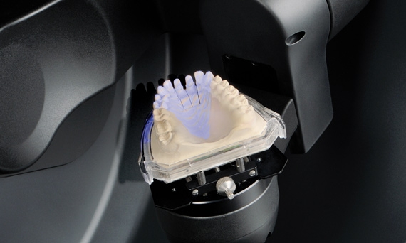
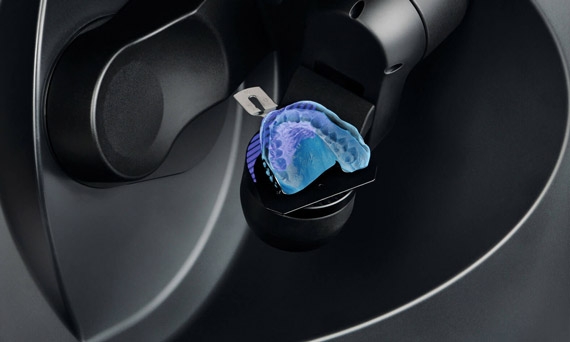
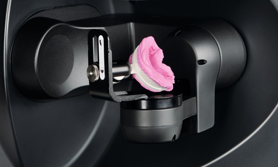
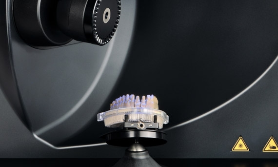
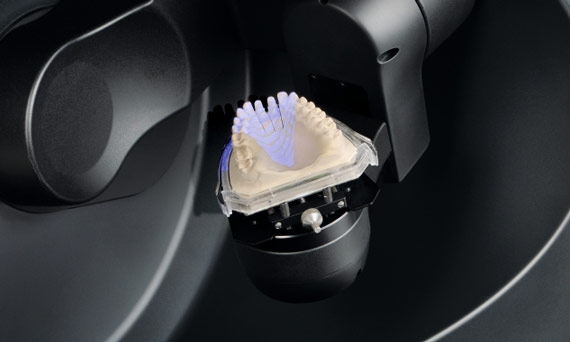
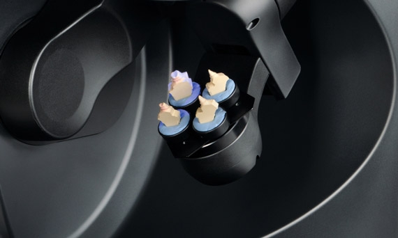
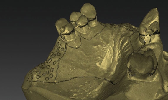
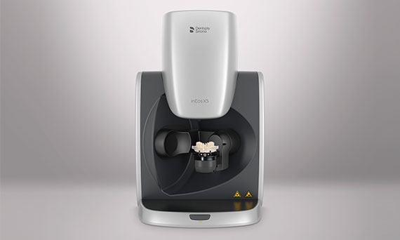
The inEos X5 was developed according to the highest quality standards for optical measuring systems. It has quickly become established on the market as the reference scanner. The inEos X5 ensures greatest accuracy for all digitization work of interest to the dental technician.

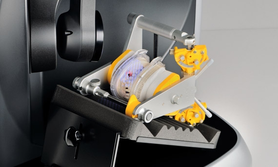
  • Intelligent scanning technology: with its robot arm, unique 5-axis scanning technology and large working area
  • Outstanding accuracy and highest depth of field
  • Clear software interface and intuitive operation result in digital models with just a few clicks
2.1 µm*

Impressive precision (0.0021 mm) as the baseline for the original situation

Reliable

All scanning tasks are carried out with extreme reliability

Open

STL data export for a free choice of design and production components

inEos X5 – Precise capture, comfortable operation, flexible function

Whether it be veneers, inlays or onlays/tabletops, or tooth or implant-supported crowns and bridges, or metal-based partial prostheses – with inEos X5 the working models can be scanned for all indications.

Precise

Detailed mirroring of real situations, from the palate to the tip of the scanbody – due to its accuracy of 2.1 µm.

Comfortable

The large operating range allows positioning of most common articulators and gives fast, unobstructed access to the scan object.

Flexible

With flexible positioning of impressions, singular or articulated models, inEos X5 guarantees the greatest possible freedom for dental applications.

Thanks to its accuracy and the high pivoting range, the inEos X5 can usually capture all scan areas in the automatic mode – even in cases with very large undercuts and strongly tilted abutment teeth.

Ingo Raschert, dental technician

Special optics

Based on digital stripe light projection with blue light, the camera optics are produced according to strict quality standards, enabling inEos X5 to scan all situations with the greatest reliability. All optical components of inEos X5 are designed and produced specifically for dental applications.

inEos X5 in action


Applications

Scan technology – Made in Germany. All inEos X5 components were specially developed in Germany for dental applications according to strict quality standards. The inEos X5 ensures precise digital registration of all indication types with its robot arm, unique 5-axis scanning technology combined with large working area.

Impression scan

The inEos X5 scans many different shapes and sizes of impression trays with no difficulty.

Triple tray scan

The lower and upper jaw together with the bite registration can be scanned from a triple-tray impression tray for smaller jobs.

Manual scanning

Smaller jobs with only a few prepared teeth can be scanned quickly and efficiently in manual mode. Open scanner The model data collected with inEos X5.

Rapid scanning

Full-arch models are digitized in less than 60 seconds thanks to the large scanning field.

Multi-die scanning

Up to four prepared dies are scanned automatically and inserted into the digital model with no manual interaction.

Texture scan

Marks on the model are detected for visual support, for example in partial-denture design using the inLab CAD software.

Wide operating range

Allows the positioning of most common articulators and gives fast, unobstructed access to the scan object.

* The accuracy of the results of the inEos X5 with inLab CAD SW 16.0 was verified according to DIN EN ISO 12836.2015. The demonstrated accuracy on standard “bridge” test specimens was at 2.1 ± 2.8 μm, and on standard “inlay” test specimens 1.3 ± 0.4 μm.


inEos X5

  Data
Dimensions (W x H x D) in mm
474 x 735 x 460
Weight
40 kg
Mains voltage
100–240 V
Mains frequency
47–63 Hz
Power consumption
150 W
Scanning technique
Digital light stripe projection
Scan material
All commonly used dental model casts (except strongly absorbent, reflecting or transparent materials) 
Interfaces
USB 2.0
Network connection
via scanner PC: LAN / WiFi (optional)

Discover the inEos X5

Implant treatments

inEos X5 – the reliable partner for implant treatments

inLab Tips & Tricks for users

Here you will find useful tips about working with the inEos X5 scanner. Download the PDF to learn more.


inLab Downloads

Here you find useful downloads for inLab users.


Technical documentation