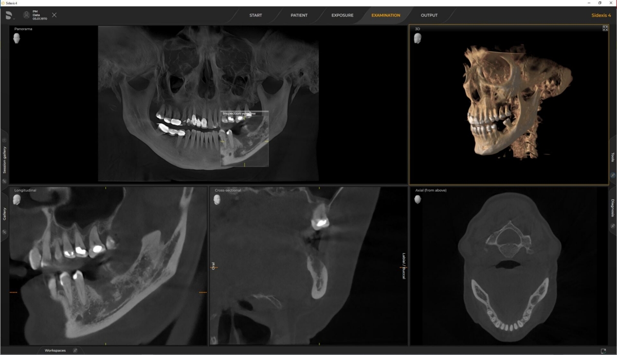
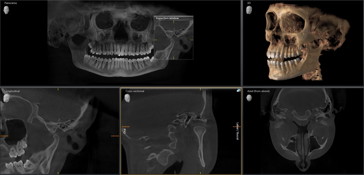
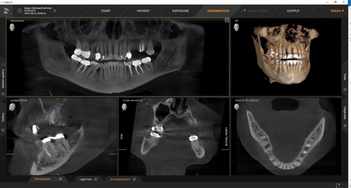
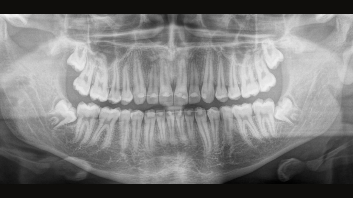
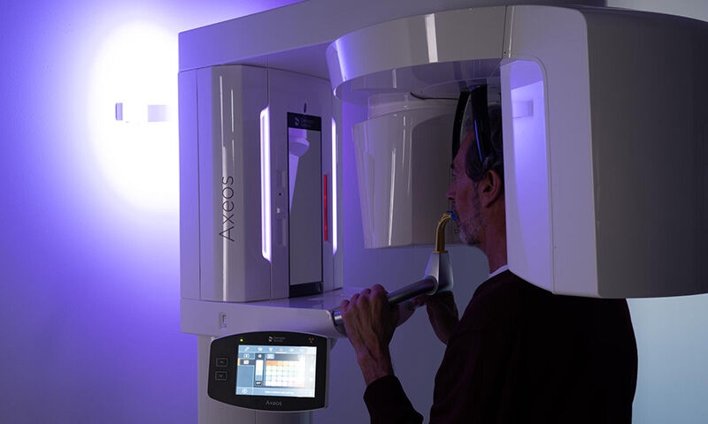
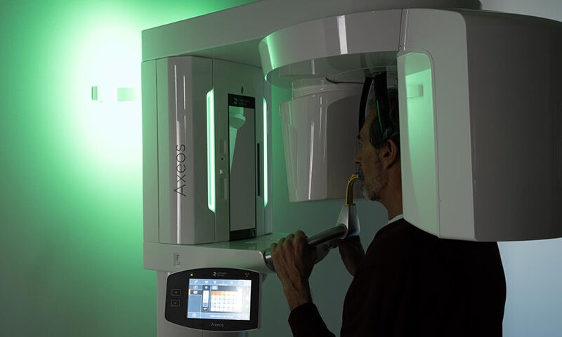
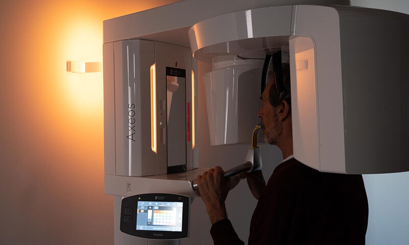
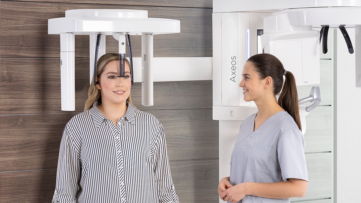
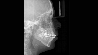
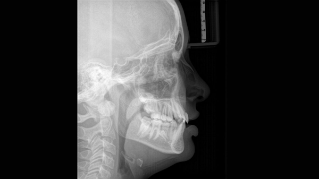
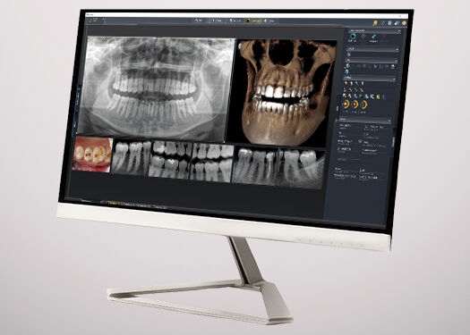
Reliable Diagnosis vs. Radiation Dosage
When trying to capture a successful medical or dental X-ray there is an ongoing fundamental conflict. On the one hand, you want to do whatever it takes to acheive maximum image quality, on the other hand, the radiation dose should be as low as reasonably achievable for the patient. At Dentsply Sirona we are dedicated to offering products that ensure exceptional image quality while supporting safe and ethical practice. When developing our products, we observe the internationally valid ALARA principle (as low as reasonably achievable).
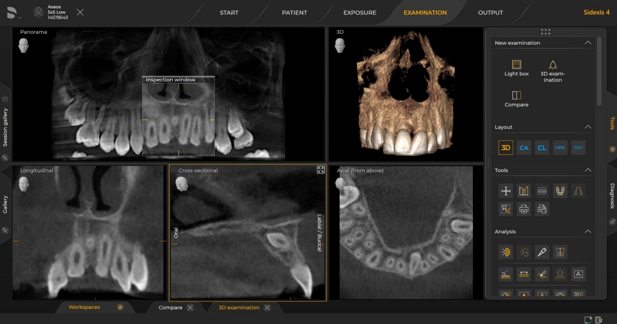
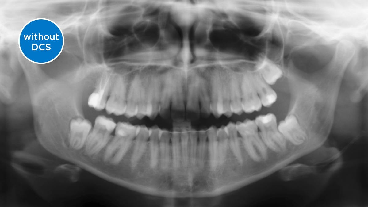
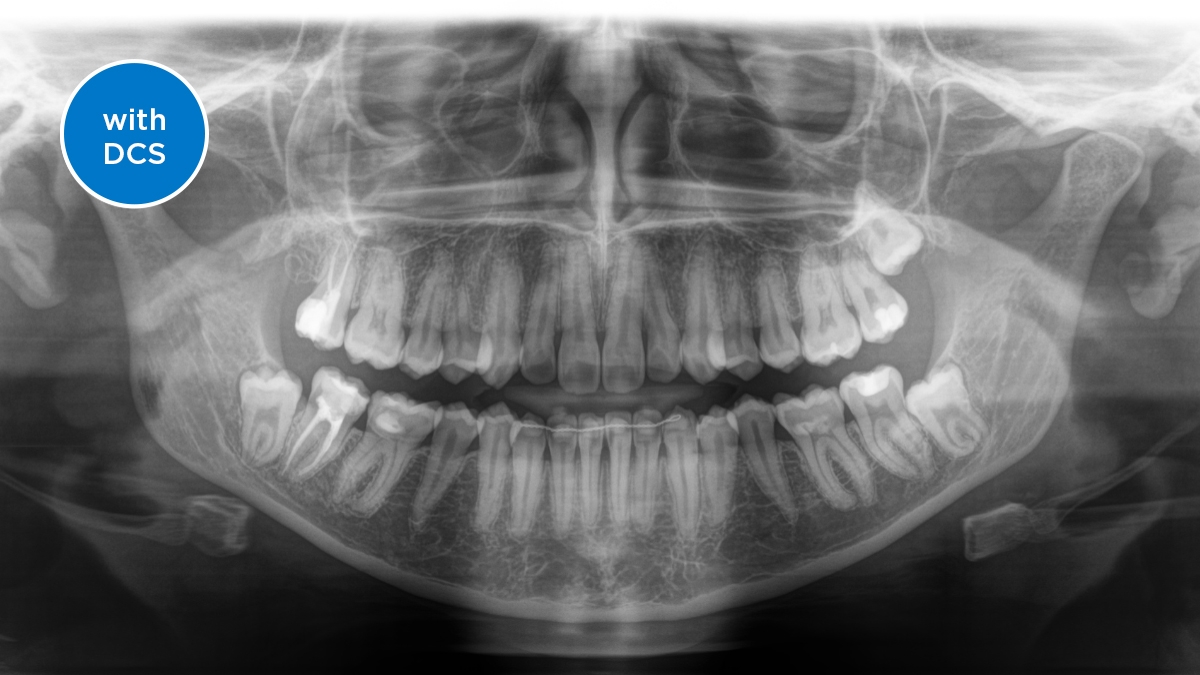
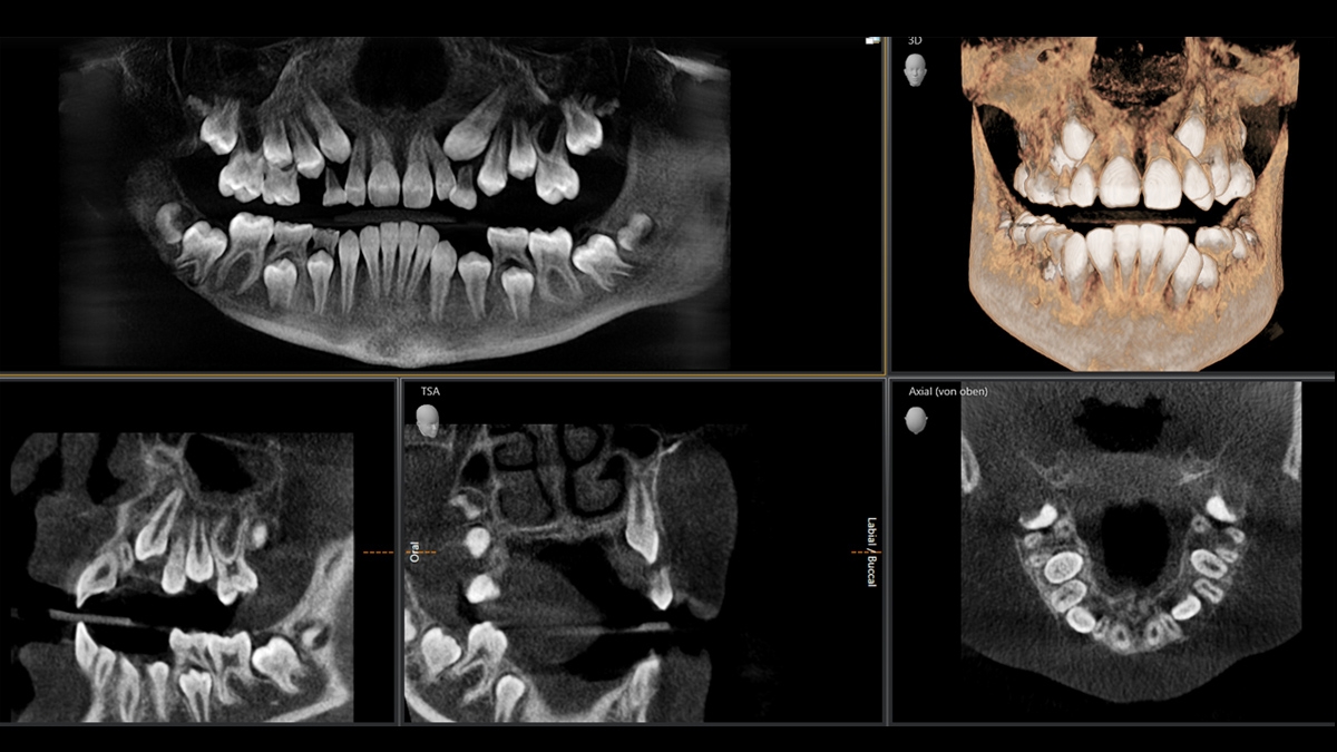
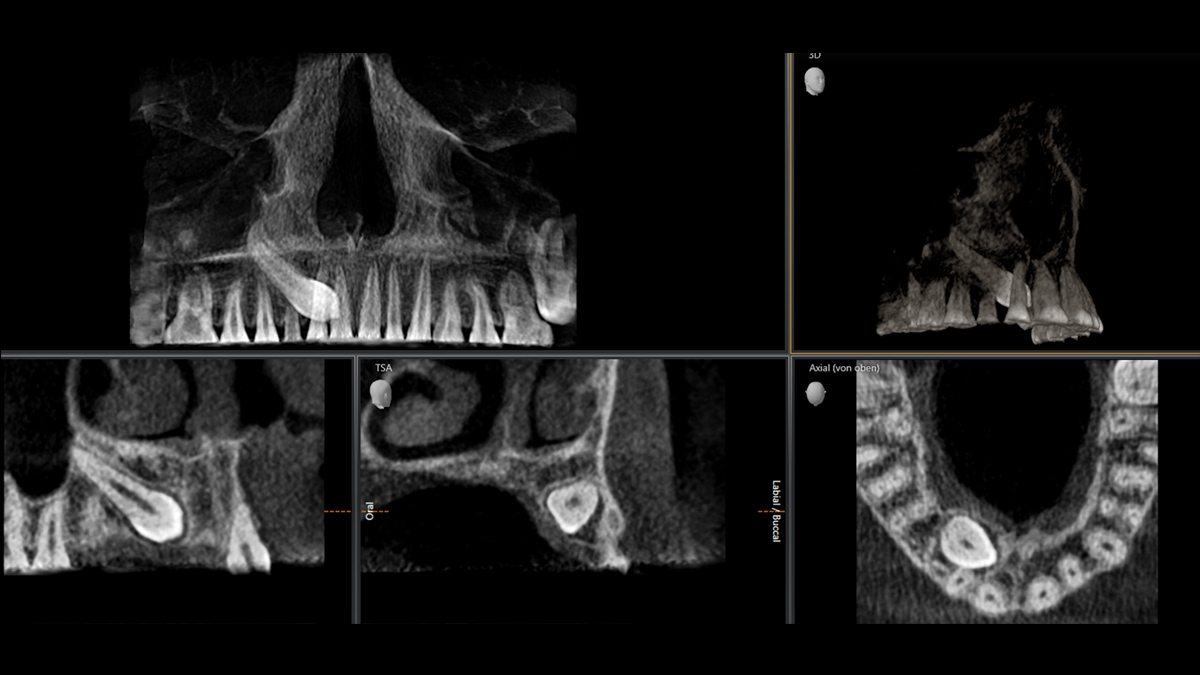
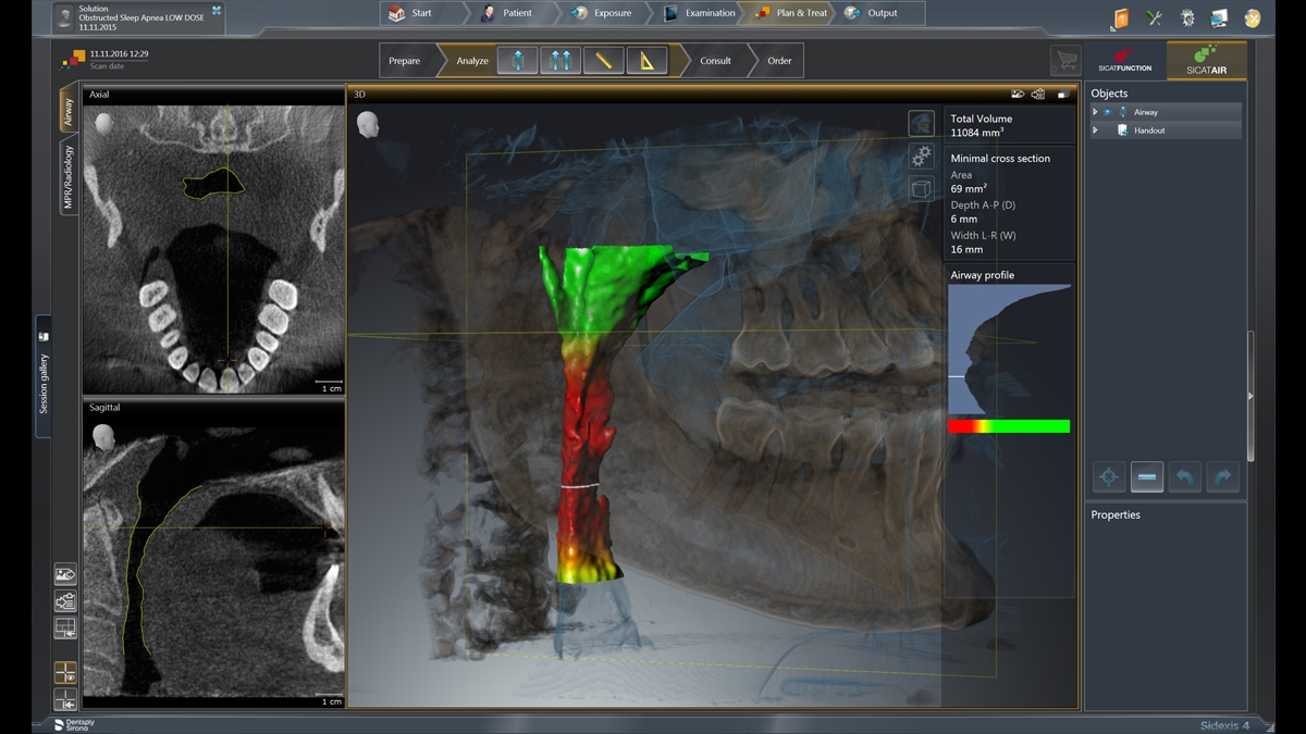
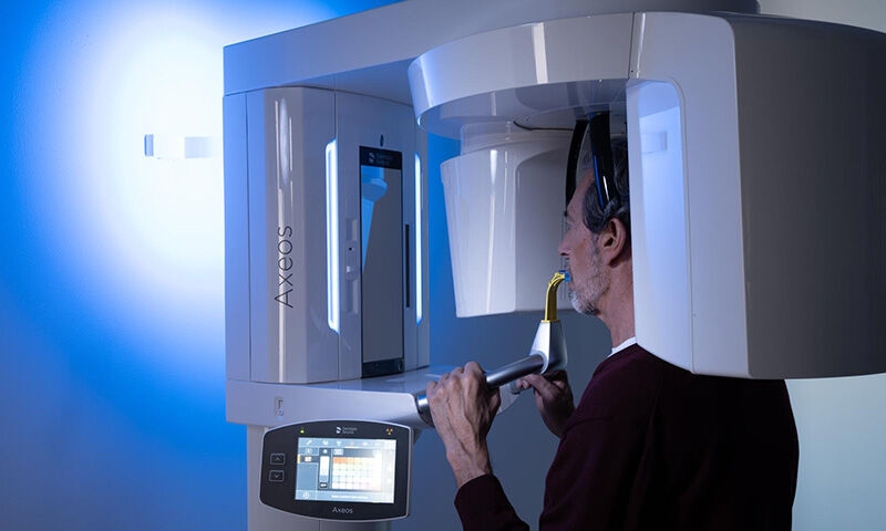
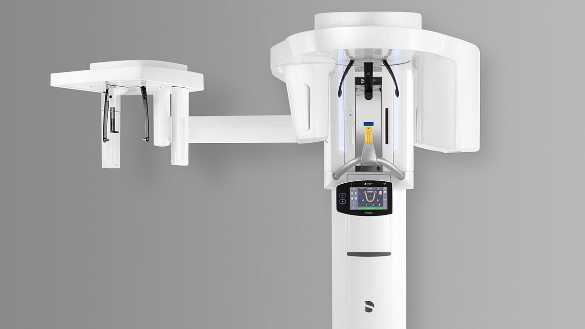
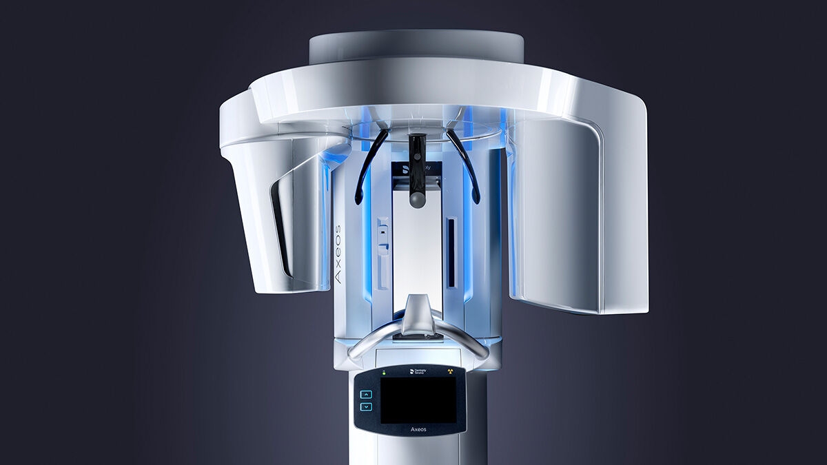
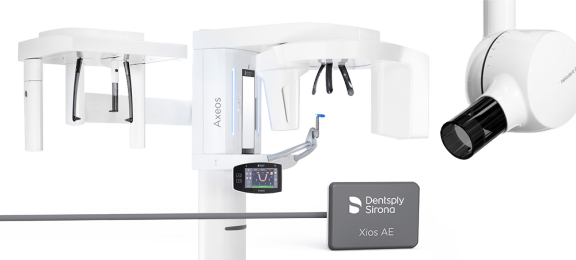
Intelligent Low Dose
Thanks to the optimised low dose mode with a dedicated filter, the imaging of dense structures, such as bones, is possible at a greatly reduced dose. This makes Intelligent Low Dose an attractive and efficient option for many clinical situations. Whether in orthodontics or implantology - with Dentsply Sirona solutions you will find the optimum setting for every case.
- Indication based - 3D low dose is suitable for a variety of clinical situations
- Dedicated filter - In contrast to conventional low dose approaches, Dentsply Sirona's Intelligent Low Dose not only lowers the mA but also applies a dedicated copper filter. This enables good visibility of dense structures (e.g. bone) despite the low dose
- Minimise dose exposure: With the 3D low dose mode you get a 3D image in the dose range of a 2D X-ray