Digital Dental X-Rays

image-comparison grip-lines arrow-top-right zip arrow-right video minus arrow-back text shop twitter file home quick-order arrow-left youtube search arrow-down check all filter site_references image Slice 1 zoom 🎬 icons / _ icon size / _ download-queue Created with Sketch. chevron-down arrow-right picture pdf podcast exe plus hotline close xxx-1 burger clipboard linked-in ppt Artboard 🎬 icons / _ icon size / _ download-queue_add Created with Sketch. Layer 1 Created by potrace 1.15, written by Peter Selinger 2001-2017 My Account press_releases Created with Sketch. PRESS ds-instruments ds-preventive ds-implants ds-endodontics Cart presentation Created with Sketch. DS-Preventive-Icon-Grey video Created with Sketch. other Created with Sketch. text_piece Created with Sketch. promotion Created with Sketch. Spare Parts ds-imaging-systems case_study Created with Sketch. ds-treatment-centers broschure Created with Sketch. ds-prosthetics ds-restorative video Created with Sketch. flyer Created with Sketch. whitepaper Created with Sketch. article Created with Sketch. image Created with Sketch. Service ds-cad-cam Artboard Quick Order Dentist Chair study Created with Sketch. ds-orthodontics ds-implants ds-endodontics ds-cerec digital-scan grinder ds-diagnosis END-Website-Icons-Shaping-Cleaning-Obturation step_1 analyse ds-abutment-placement more-quality initiative scan more-quality ds-polishing digital ds-irrigation ds-treatment-centers Test END-Website-Icons-Shaping-Cleaning-Obturation treatment ds-restorative ds-implant-planning realistic construct book ds-planning-impression END-Website-Icons-Shaping-Cleaning-Obturation END-Website-Icons-Shaping-Cleaning-Obturation mirror anwender ds-preventive planning-2 ds-obturation ds-cad-cam 04 ds-implant-placement ds-retention-protection ds-anesthetics restauration form ds-inlab oven CORP-icon-grey-screen-tooth-cavity-pulpa robot plan place ds-orthodontics PDF-Formular_ganzneu PDF overlay ds-bone-regeneration ds-treatment-preparation vorher_nachher ds-maging-systems wifi END-Website-Icons-Shaping-Cleaning-Obturation omnicam PDF-Formular_neu_vonGünther treatment consulting planning comparison ds-set-up ds-prosthetics support step_3 ds-tooth-preparation ds-treatments ds-scaling step_2 ds-instruments software quote-english quote-french quote-text-german Created with Sketch. upload Created with Sketch. version Created with Sketch. globe EndoEquipmentGreypng Imaging Grey 2svg EndoGlideShapingGreypng irrigation blue Imaging 1svg Imaging 2svg DS-Preventive-Icon-Grey Final_Implants_Category_Implant-Systems Access Blue obturation blue restoration restoration grey Access Grey treatment EndoEquipmentBluepng irrigation grey Diagnosis primeprint-grey Hub Bluesvg Final_Implants_Procedure_Single-tooth Diagnsis blue Imaging blue EndoRetretmentBluepng restoration blue Final_Implants_Procedure_Edentulous-therapy Final_Implants_Procedure_Hard-and-soft-tissue-management obtruation grey endo grey treatment blue Imaging blue 1svg Imaging grey EndoGlideShapingBluepng EndoRetretmentGreypng polishing Hubsvg Final_Implants_Category_Implant-Dentistry polishing blue Diagnosis Grey Cerec Bluesvg Imaging Blue 2svg treatment grey Hub Greysvg Scaling Cerec Greysvg endo blue polishing grey search Imaging Grey 1svg scaling blue Cerecsvg DS Points Logo Imaging scaling grey

Digital Dental X-Rays with Market-Leading Accuracy and Precision

Dentsply Sirona delivers unparalleled diagnostic support with a comprehensive line of digital x-ray systems with market-leading accuracy and precision. With more than 120 years of imaging experience, our workflow solutions are made with the practitioner in mind – making the diagnostic process safer and more efficient. Optional technology, sensors, and add-ons make the modular design of our x-ray units easy to integrate and expand with your practice.

The Digital Advantage

  • Healthy:  Lower radiation exposure
  • Precise:  Outstanding image quality produces brilliant images
  • Efficient:  Quicker processing and efficient data management
  • Integrated:  Mobile solutions and integrated technology to connect data systems
  • Accurate:  A variety of analysis tools for simplified, accurate diagnosis
  • Educational:  Visual images readily available for patient education in vivid detail

Axeos Imaging System

Axeos 2D/3D system was designed for the clinician who offers a wide range of treatments. This system combines high-quality images with enhanced comfort for the patient.

Our Direct Conversion Sensor combines very sharp images with low radiation doses. For you, this means clinical confidence based on four advantages offered by all our 3D solutions:

  1. According to the clinical circumstance, you have access to a volumes per click range of Ø5 cm x 5.5 cm to Ø17 cm x 13 cm.
  2. Ability to obtain a high resolution, low-noise image in HD mode with 1400 single images being captured during one circulation.
  3. Using the innovative algorithms of MARS, you can automatically identify and minimize metal artifacts.
  4. Our Intelligent Low Dose mode is beneficial for acquiring superior images of dense structures.

Dentsply Sirona is committed to providing high-quality imaging while minimizing patients’ radiation exposure by offering products such as this CBCT scanner that delivers sharp, quality images while keeping the radiation dose as low as is reasonably achievable.

Orthophos Family

The Orthophos family for extraoral imaging offers the right solution for every practice – from entry into digital radiography to specialty services. Orthophos imaging units are simple and intuitive to operate, with smart technology to assist with patient positioning and image quality. Modular options and custom controls allow the practitioner to quickly incorporate the imaging system into current workflows and expand the technology to grow with the practice and clinical offerings. 

Versatile & Safe – Low Dose 

The Orthophos family has been developed according to the ALARA principle to allow for the best X-ray images with the lowest radiation dose. All programs and capture parameters are tailored to the specific diagnostic tasks, such as pediatric panoramic program with reduced image dimensions and a QuickScan function with faster exposures.

Extra Oral Bitewing

With all Orthophos models, you can use the bitewing function to create extraoral bitewing images with an optimized curve for the posterior tooth region. With the image field selection, you can focus on the area of interest.

Optimal Stability

Stable patient positioning prevents motion blur. The motorized 3-point head fixation and sturdy handles give your patient the necessary support. The integrated temple width measurement automatically ensures a patient-specific orbit. Unnecessary downtime can be reduced by the automatic opening of the temple support for a successful X-ray outcome.

Entry-Level Imaging

Orthophos E

The solid entry-level unit for basic diagnostics, Orthophos E provides a smooth entrance into the world of digital imaging. The 2D imaging unit offers easy and thoughtful operation with its MultiPad Control panel.  Achieve reliable image quality with the CsI sensor and sophisticated technical support with Sidexis 4 Software.

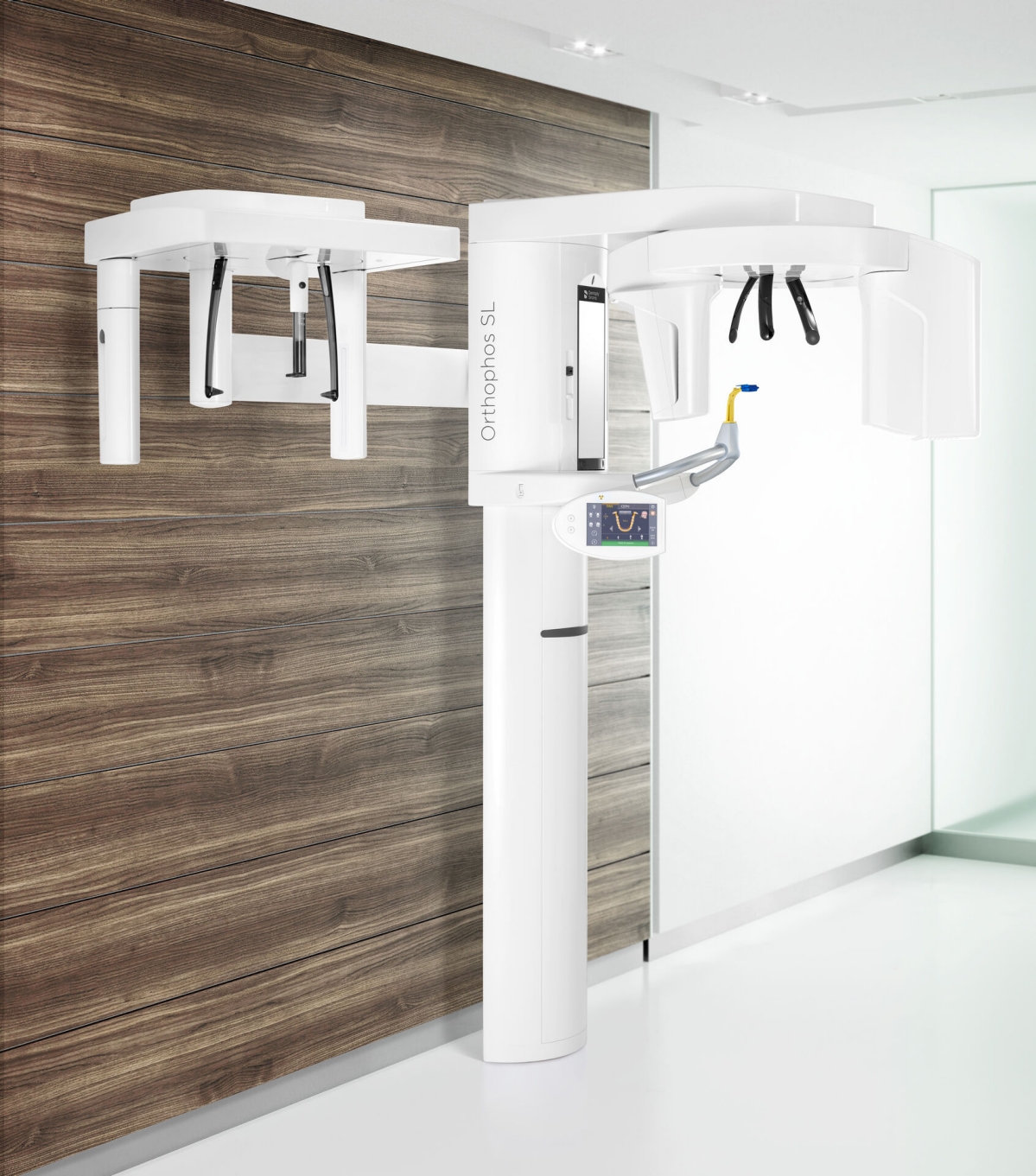
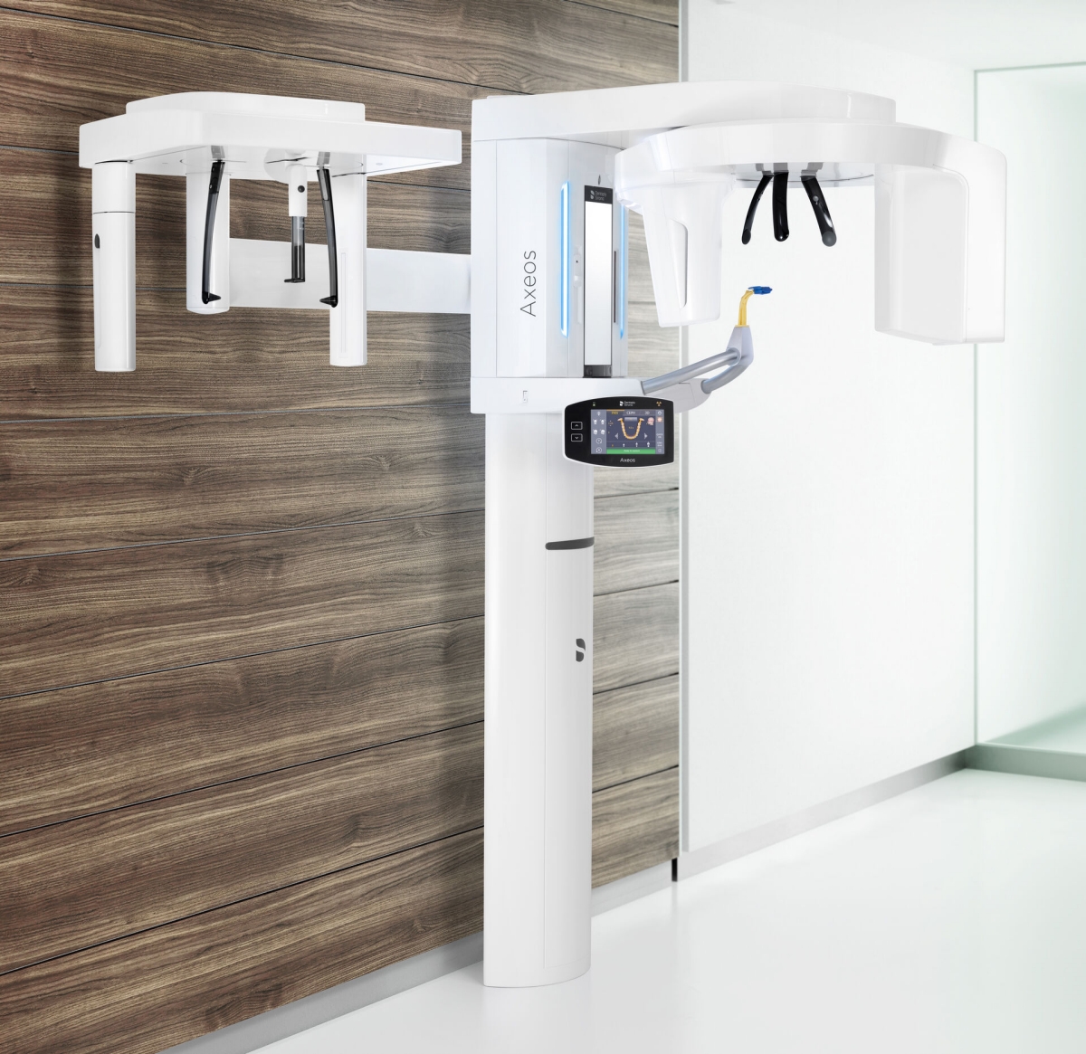
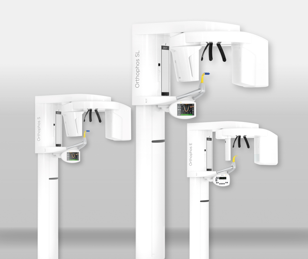
2D/3D Imaging Systems

Intuitive Operation 

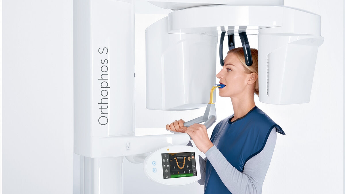
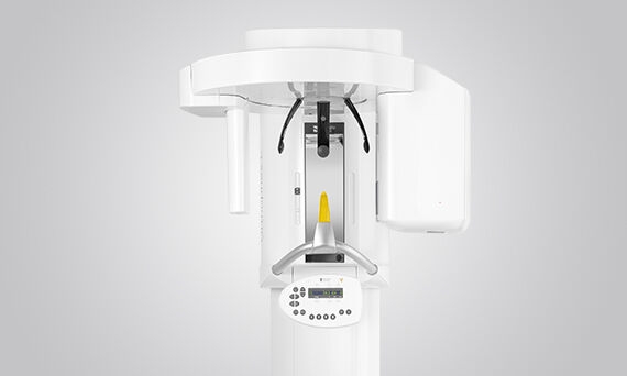
The EasyPad operating panel swivels and tilts for optimal access, with clearly marked buttons and symbols for quick navigation and fine-tuning. 

Patented Occlusal Bite Block

The patented occlusal bite block automatically establishes the correct inclination of the patient's head, providing reproducible and consistent patient positioning. The motorized three-point head fixation and stable handles give your patients the necessary support. At the same time the integrated temple width measurement ensures an orbit specific to each patient. This stability prevents motion blurring.

Autofocus

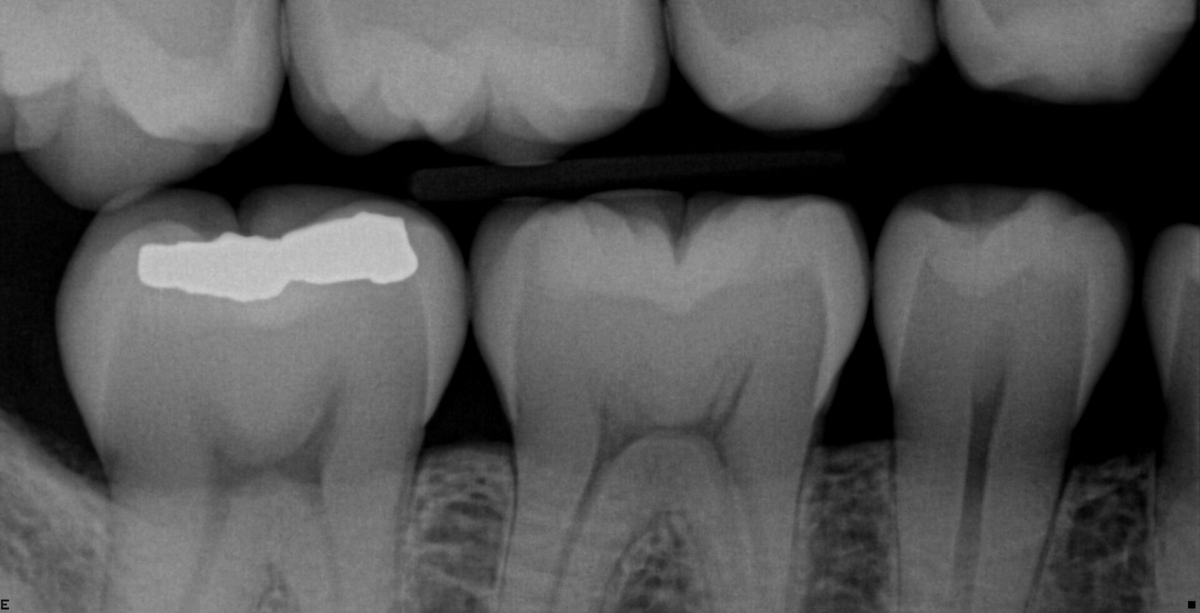
To achieve a sharp panoramic X-ray image in high definition, the right focus is essential. The jaw must be in the sharp image layer of the device. For this, the Orthophos creates several thousand individual images in one rotation and automatically recognizes the areas in which the jaw is optimally positioned. These are displayed in an overall sharp image – without any manual intermediate steps.

Orthophos S

The Orthophos S is a high-quality X-ray unit with a comprehensive range of services optimized for everyday tasks. Whether as a 2D device, or incorporating the 3D module, the Orthophos S is a reliable partner with automatic patient positioning with its patented occlusal bite block. The CsI Plus sensor with autofocus function produces sharp images even in anatomically difficult cases. For use in orthodontics, the Orthophos S is also available with an optional ceph arm that can be retrofitted at any time.

Orthophos SL

The premium 2D/3D high-end unit for practices with a keen understanding of the latest technologies. The integrated Direct Conversion Sensor (DCS) and Sharp Layer technology redefines the standard of panoramic imaging, providing exceptional performance even in difficult cases. Orthophos SL guarantees maximum ease of use through automatic positioning, intuitive operation with the EasyPad and an individually adjustable ambient light for an exclusive look and feel. For bitewing, sinus or ceph images, left or right ceph arms are optional and can be retrofitted at any time. 

Direct Conversion Sensor (DCS) Technology

DCS Technology converts X-rays directly into electrical signals. Unlike conventional systems, there are no signal losses due to light conversion. The improved image information output delivers images with stunning sharpness, redefining the standard of panoramic imaging. The process generates optimal yield of X-ray radiation, delivering precision even with a low radiation dose.

Sharp Layer Technology

Interactive SL allows you to define an image detail of your choice to focus in on lingual/buccal objects after scanning, determining position without corrective scans.

3D Dental X-Ray

Dental 3D Imaging

The level of data delivered from a 3D image is a revolutionary force in dental diagnostics. The ability to visualize overlaid teeth, unexpected canals, and hidden roots elevates the accuracy and impact of a patient’s diagnosis. Enhancing understanding and confidence positively influences patient acceptance and treatment success.

Orthophos S and SL systems can be upgraded with 3D capabilities at any time, allowing these imaging units to match your level of services and develop with your practice. With these two 3D models, you can choose on a case-by-case basis whether to use high-resolution volumes for fine structures (HD) or a low-dose filter for dense structures.

Volume

When it comes to volume size, dose, and image quality, every clinical case demands individual requirements. Choose the appropriate volume for your needs: From the focu­sed Ø 5 cm x 5.5 cm volume to the Ø 11 cm x 10 cm volume, which displays the wisdom teeth and upper respiratory tract.

Metal Artifact Reduction Software (MARS)

Metal artifacts are a challenge in 3D imaging. Radiopaque objects create shadowing and streaking effects during the three-dimensional reconstruction, interfering with the findings. MARS automatically reduces metal artifacts and facilitates the diagnosis.

High Definition (HD) Mode

Standard Definition Mode (SD) provides basic clinical information needed for a diagnosis. Some clinical cases require a higher quality image with better visibility of fine structures for treatment planning and implementation. The HD mode records up to 800 individual images during one rotation and merges them into a low-noise, 3D volume with high resolution of up to 80 μm. This mode guarantees a faster and safer diagnosis within the recorded volume.

Low Dose Mode

The optimized Low Dose Mode with a dedicated filter allows for the imaging of dense structures, like bone, at a greatly reduced dose. This makes Low Dose an efficient option for many clinical tasks – especially for those in orthodontics or implantology. With 3D imaging capabilities, patients no longer need a referral for a CBCT scan, streamlining the process and expanding your service offerings.

Other Imaging Solutions

Sidexis Software

Sidexis 4 Software brings your brilliant images to life, giving you everything you need in a glance. Deliver your proposal in high-definition, reviewing 2D and 3D images, photographs, and video in side-by-side comparison for a comprehensive treatment overview.

Review patient history with a virtual timeline, giving visual support to treatment options and increasing patient understanding and acceptance. With the Sidexis iX App for iPad, take your images mobile and make your iPad a comfortable advisory tool.

Intraoral Sensors & X-Ray Generator

Dentsply Sirona delivers comprehensive diagnostic solutions that elevate the potential of your digital x-ray system. By further enhancing image clarity, custom settings, and versatility, you can take your diagnostic workflow from advanced to exceptional.

Schick 33

Schick 33 Intraoral Sensors bring an unprecedented level of clarity, sharpness, and detail to digital radiography. The Schick 33 sensors integrate seamlessly with the Dentsply Sirona X-ray units and software to deliver better, safer, and faster diagnostics.

Clinicians can rely on Schick’s superior image quality to meet their diagnostic needs, featuring bold bone trabeculation, crisp lamina dura, and a clear dental enamel junction. With three sensor sizes, multiple cables lengths, and limitless enhancement capabilities, Schick 33 is designed to suit your patient's needs, as well as your practice and personal preference.

Integrated Technology

Building on 25 years of digital intraoral radiography, Schick 33 uses Dentsply Sirona’s 2nd generation CMOS-APS imaging technology enhanced by a dynamic software solution – Sidexis 4.  The seamless integration allows the data rich images to be immediately available on screen in high-definition.

High Image Quality

Image clarity is the most cited technical factor for quality imaging. Outstanding image resolution is essential for defining edges, identifying caries and other defects. At 33 lp/mm theoretical resolution, Schick 33 delivers industry-leading image resolution and quality.

The superb image quality provides an accurate and compelling visual reference when discussing treatment options and benefits with your patients. The visual advantages can lead to a more comprehensive diagnosis, higher clinical confidence, and greater patient acceptance.

Image Optimization

With Schick 33 sensors you have the option of five different filters: General Dentistry, Endodontics, Periodontics, Restorative, and Hygiene. These clinically specific views are designed to optimize your diagnosis, enhancing the image to suit your needs.

Further adjust your diagnostic settings with the dynamic sharpening slider, individually controlling sharpness, brightness, and contrast. Configure your display for optimal viewing and save the preset for future use, ensuring a consistently efficient diagnostic approach. 

Robust & Durable

Detachable cables of differing lengths allow Schick 33 sensors to accommodate any size operatory. The simple cable exchange makes installation and adjustments quick and easy. The robust sensor interface gives you an extremely durable USB connection, while the Kevlar reinforced, silicon-coated cables help keep your sensor up and running. 

Heliodent Plus

Heliodent Plus is the latest generation of intraoral x-ray generator designed for optimum image quality in digital radiography. With 0.4 mm focal spot and vibration-free components, Heliodent Plus offers smart design for stable positioning and ultimate image quality.

The Heliodent Plus offers a broad range of mounting options to allow for optimum configuration in each treatment room. The equipment has been designed to support your treatment style and allow you to work exactly how you’re comfortable. An intuitive user display with pre-programed settings and easy service capabilities help deliver greater clinician control for day-to-day practice workflow.

Installation Options

  • Wall Model
  • Integrated Treatment Center
  • Mobile Unit
  • Ceiling Mount

Operation Options

  • Remote Control
  • Remote Timer
  • Manual Release

Contact us for more information

Find out more about the imaging systems from Dentsply Sirona and request information on intraoral imaging, software or 2D and 3D imaging technology.