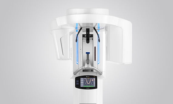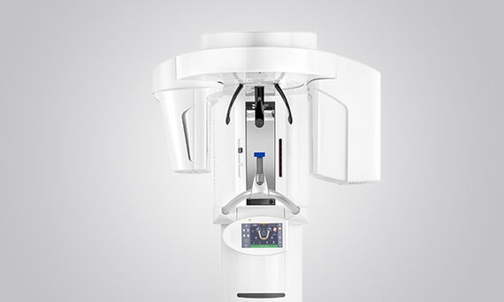
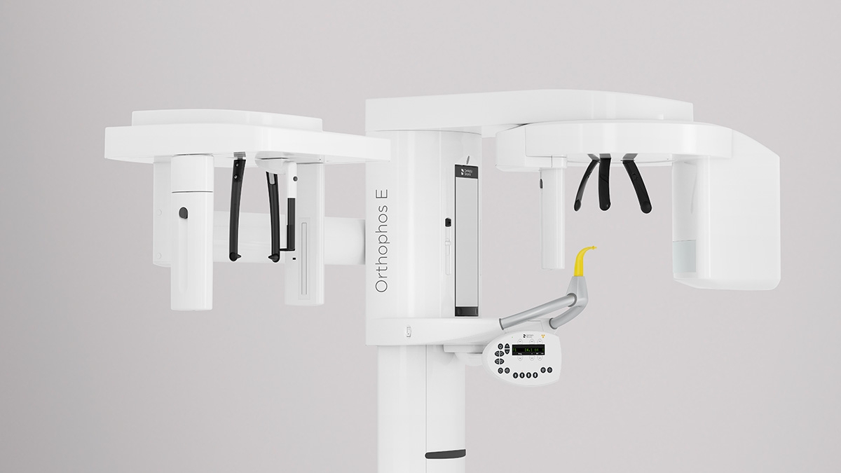
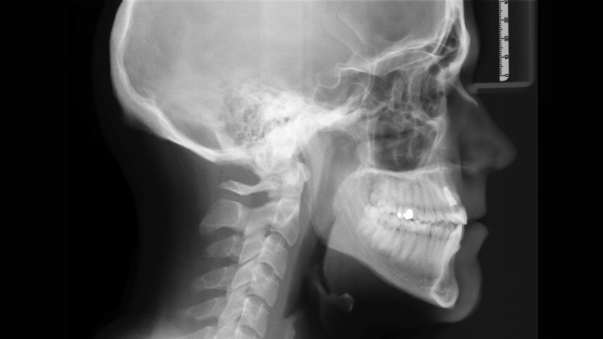
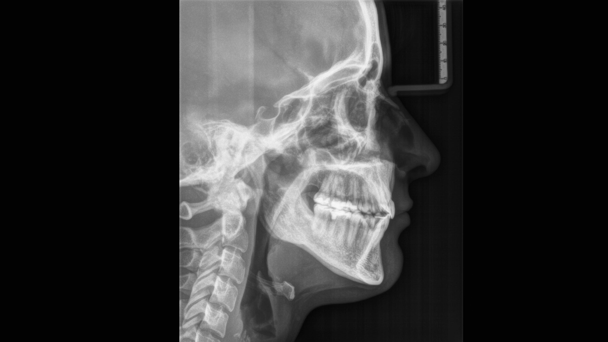
Proven patient positioning
Only 2 light localizers are required for ideal positioning in the sharp slice. The motorized forehead and temple supports fixate the patient's head and prevent motion blurring.
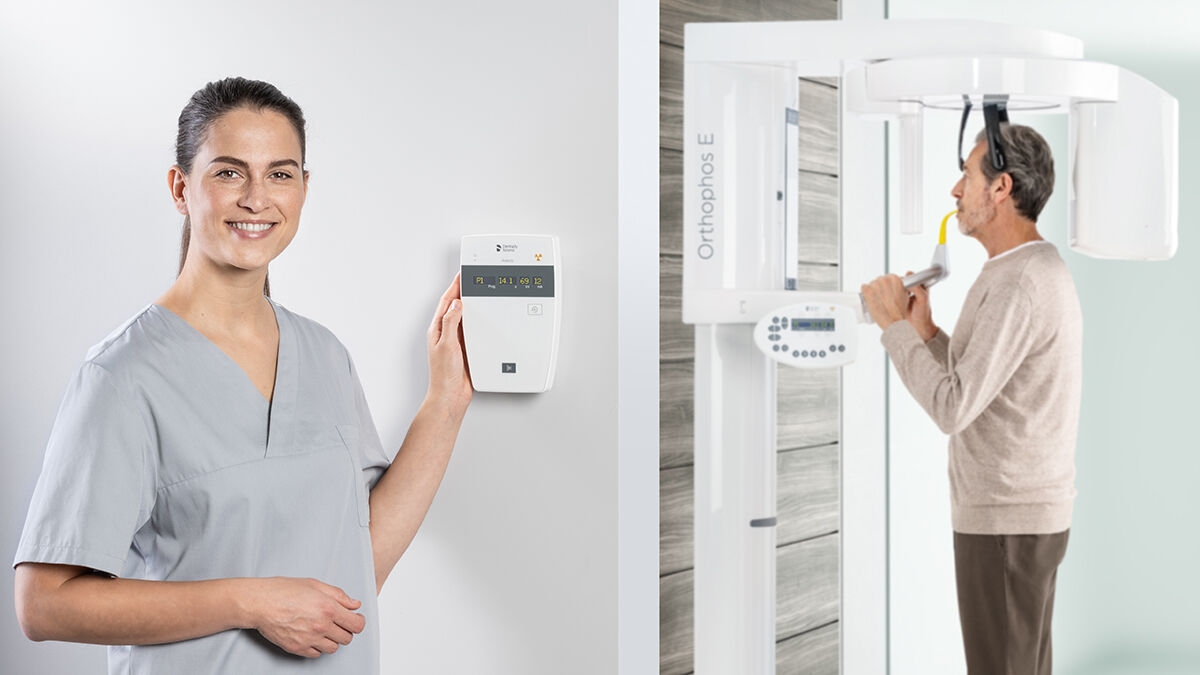
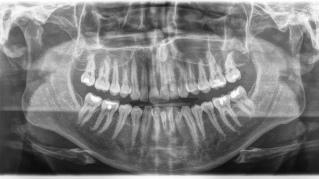
The sound 2D x-ray unit providing a smooth entrance into the world of digital imaging. Thanks to CsI sensor technology and its easy-to-use interface, you are ensured reliable diagnostics every time. The cephalometric option also makes the Orthophos E a reliable partner for orthodontics. Enrich your practice with a wide range of services that are only possible with digital imaging.
The Orthophos E is the go-to solution for general dentists and orthodontists. Diagnostic accuracy is made possible by the many advantages you get when choosing digital imaging.
Thanks to the 2D-CsI Plus sensor and reliable image quality
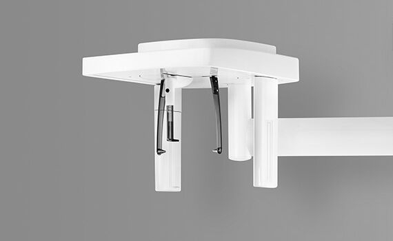
With motorized temple and forehead support, automatic temple width measurement, light localizers and sturdy handles
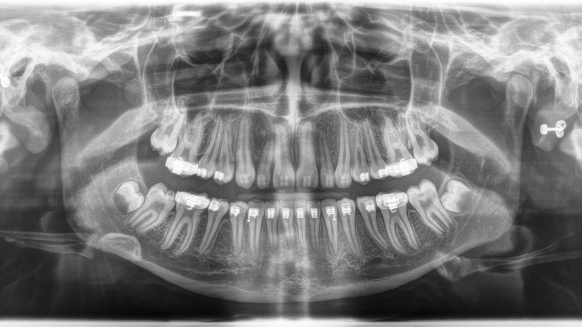
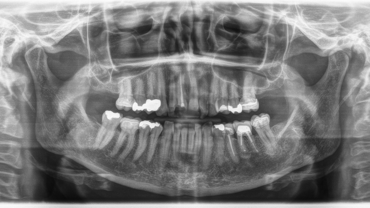
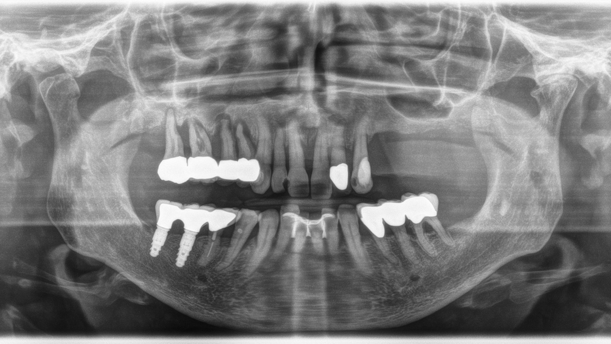
For basic diagnostics in 2D
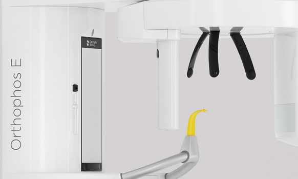
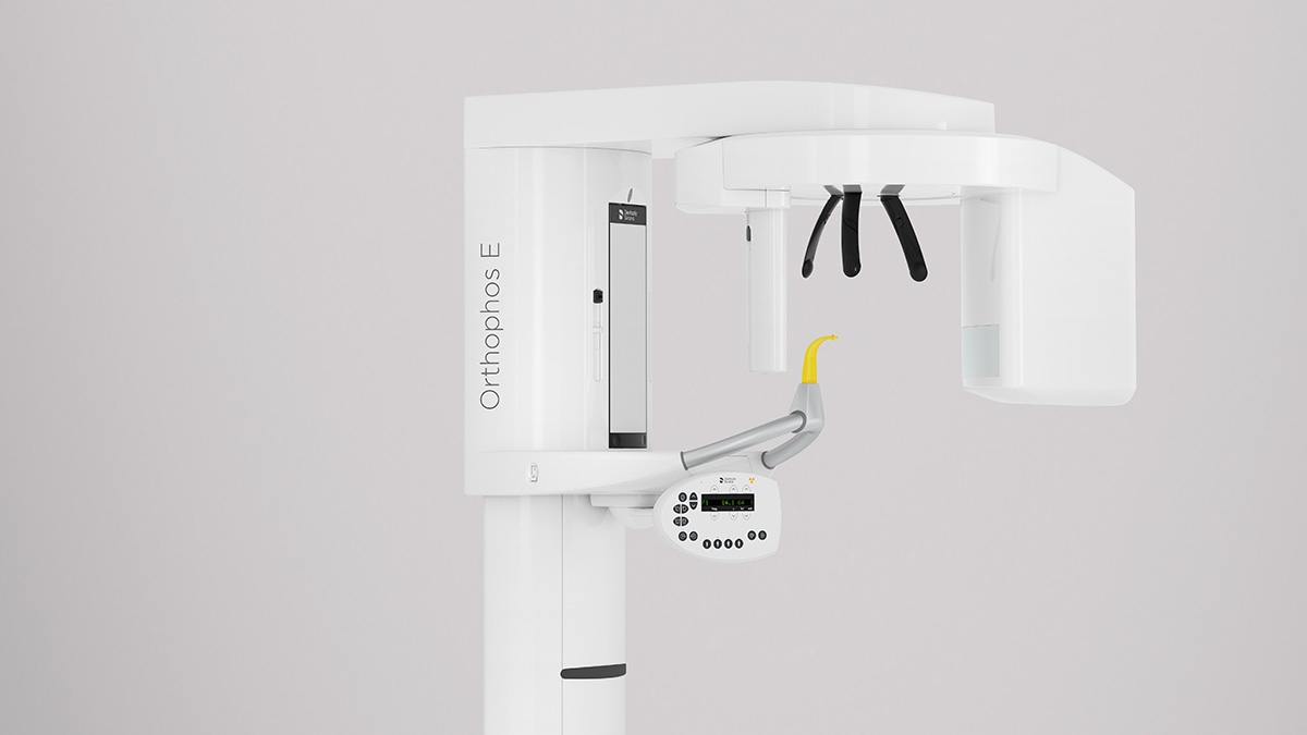
With the MultiPad control panel
With optional left Ceph
Given indications
Given indications
Given indications

Given indications


| Programme | Orthophos SL 2D | Orthophos S 2D | Orthophos E |
|---|---|---|---|
| Standard panorama image |
P1, P2, P10 |
P1, P2, P10 |
P1, P10 |
| Image detail left side or right side |
P1, P1A, P1C P2, P2A, P2CP10, P10A, P10C BW1 |
P1, P1A, P1C P2, P2A, P2CP10, P10A, P10C BW1 |
P1L, P1R |
| Image detail individual quadrants |
P1, P1A, P1C P2, P2A, P2CP10, P10A, P10C |
P1, P1A, P1C P2, P2A, P2CP10, P10A, P10C |
- |
| Image detail upper or lower jaw |
P1, P1A, P1C P2, P2A, P2CP10, P10A, P10C, P12 |
P1, P1A, P1C P2, P2A, P2CP10, P10A, P10C, P12 |
- |
| Constant magnification |
P1C, P2C, P10C |
P1C, P2C, P10C |
P1C |
| Artifact-reduced |
P1A, P2A, P10A |
P1A, P2A, P10A |
P1A |
| Thick layer front |
P12 |
P12 |
P12 |
| Sinusoidal images |
S1, S3 |
S1, S3 |
S1 |
| Multislice in posterior tooth |
- | - | MS1 |
| Mandibular joint |
TM1.1, TM1.2, TM3 |
TM1.1, TM1.2, TM3 |
TM1.1, TM1.2 |
| Bitewing image |
BW1, BW2 |
BW1, BW2 |
BW1 |
| Ceph (optional) |
C1, C2, C3, C3F, C4 |
C1, C2, C3, C3F, C4 |
C1, C2, C3, C3F, C4 |
Only 2 light localizers are required for ideal positioning in the sharp slice. The motorized forehead and temple supports fixate the patient's head and prevent motion blurring.
| Programme | Orthophos SL 2D | Orthophos S 2D | Orthophos E |
|---|---|---|---|
| Standard panorama image |
P1, P2, P10 |
P1, P2, P10 |
P1, P10 |
| Image detail left side or right side |
P1, P1A, P1C P2, P2A, P2CP10, P10A, P10C BW1 |
P1, P1A, P1C P2, P2A, P2CP10, P10A, P10C BW1 |
P1L, P1R |
| Image detail individual quadrants |
P1, P1A, P1C P2, P2A, P2CP10, P10A, P10C |
P1, P1A, P1C P2, P2A, P2CP10, P10A, P10C |
- |
| Image detail upper or lower jaw |
P1, P1A, P1C P2, P2A, P2CP10, P10A, P10C, P12 |
P1, P1A, P1C P2, P2A, P2CP10, P10A, P10C, P12 |
- |
| Constant magnification |
P1C, P2C, P10C |
P1C, P2C, P10C |
P1C |
| Artifact-reduced |
P1A, P2A, P10A |
P1A, P2A, P10A |
P1A |
| Thick layer front |
P12 |
P12 |
P12 |
| Sinusoidal images |
S1, S3 |
S1, S3 |
S1 |
| Multislice in posterior tooth |
- | - | MS1 |
| Mandibular joint |
TM1.1, TM1.2, TM3 |
TM1.1, TM1.2, TM3 |
TM1.1, TM1.2 |
| Bitewing image |
BW1, BW2 |
BW1, BW2 |
BW1 |
| Ceph (optional) |
C1, C2, C3, C3F, C4 |
C1, C2, C3, C3F, C4 |
- |
Orthophos E offers the capability to install a left cephalometric arm at any time, whether initially at the time of order or retrofitted later on.


| Performance features |
Orthophos SL 2D | Orthophos S 2D | Orthophos E |
|---|---|---|---|
| X-ray generator |
60 - 90 kV, 3-16mA | 60 - 90 kV, 3-16mA | 60 - 90 kV, 3-16mA |
| Panoramic exposure time |
P1: max 14,2 s P1 Quickshot: max 9,1 s |
P1: max 14,2 s P1 Quickshot: max 9,1 s |
P1: max 14,2 s |
| Radiation time Ceph |
Standard 9,4 s Quickshot 4,7 s |
Standard 9,4 s Quickshot 4,7 s |
Standard 9,4 s |
| User interface |
EasyPad | EasyPad | MultiPad |
| Patient positioning |
automatic (occlusal bite block) |
automatic (occlusal bite block) |
manual |
| Panorama technology |
DCS | CsI Plus | CsI |
| Autofocus |
yes | yes | - |
| Ceph arm (optional) |
left or right | left or right |
left |
| Ceph unit with 2 sensors |
yes |
yes |
optional |
| Quickshot |
yes |
yes |
- |
| Fields of View |
upgradeable | upgradeable |
- |
| 3D Low Dose |
upgradeable |
upgradeable |
- |
| HD mode |
upgradeable |
upgradeable |
- |
| Base |
optional |
optional |
optional |
| Wheelchair accessible |
yes | yes | yes |
| Remote control |
optional |
optional |
optional |
| Ambient Light | yes (backlight) | - | - |
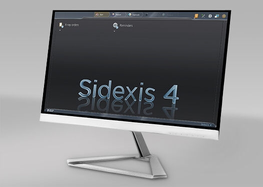
For more details please see Sidexis 4 system requirements
Dentsply Sirona 2D units work exclusively with Sidexis 4. Nevertheless data migration from Sidexis XG to Sidexis 4 is very easy. Sidexis 4 allows for the full digital experience with the latest tools.
The Orthophos SL 2D and the Orthophos S 2D allow a 3D upgrade. Axeos is a dedicated hybrid unit. The Orthophos E does not offer this option.
Find out more about our 2D and 2D/3D hybrid product range and download the extraoral X-ray family brochure here.
Find out more about Dentsply Sirona Imaging Solutions for Implantology
Find out more about Dentsply Sirona Imaging Solutions for Orthodontics
Whether hardware or software, a variety of imaging solutions are waiting for you.