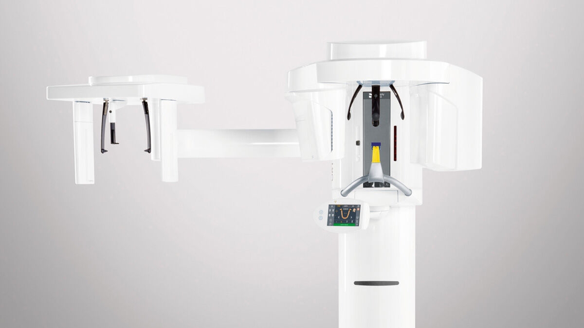
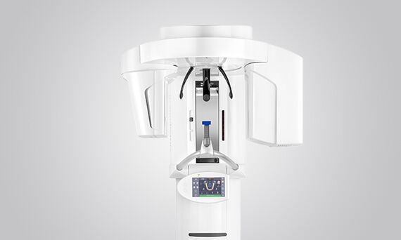
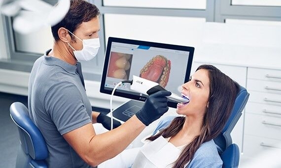
3D Dental Imaging

image-comparison grip-lines arrow-top-right zip arrow-right video minus arrow-back text shop twitter file home quick-order arrow-left youtube search arrow-down check all filter site_references image Slice 1 zoom 🎬 icons / _ icon size / _ download-queue Created with Sketch. chevron-down arrow-right picture pdf podcast exe plus hotline close xxx-1 burger clipboard linked-in ppt Artboard 🎬 icons / _ icon size / _ download-queue_add Created with Sketch. Layer 1 Created by potrace 1.15, written by Peter Selinger 2001-2017 My Account press_releases Created with Sketch. PRESS ds-instruments ds-preventive ds-implants ds-endodontics Cart presentation Created with Sketch. DS-Preventive-Icon-Grey video Created with Sketch. other Created with Sketch. text_piece Created with Sketch. promotion Created with Sketch. Spare Parts ds-imaging-systems case_study Created with Sketch. ds-treatment-centers broschure Created with Sketch. ds-prosthetics ds-restorative video Created with Sketch. flyer Created with Sketch. whitepaper Created with Sketch. article Created with Sketch. image Created with Sketch. Service ds-cad-cam Artboard Quick Order Dentist Chair study Created with Sketch. ds-orthodontics ds-implants ds-endodontics ds-cerec digital-scan grinder ds-diagnosis END-Website-Icons-Shaping-Cleaning-Obturation step_1 analyse ds-abutment-placement more-quality initiative scan more-quality ds-polishing digital ds-irrigation ds-treatment-centers Test END-Website-Icons-Shaping-Cleaning-Obturation treatment ds-restorative ds-implant-planning realistic construct book ds-planning-impression END-Website-Icons-Shaping-Cleaning-Obturation END-Website-Icons-Shaping-Cleaning-Obturation mirror anwender ds-preventive planning-2 ds-obturation ds-cad-cam 04 ds-implant-placement ds-retention-protection ds-anesthetics restauration form ds-inlab oven CORP-icon-grey-screen-tooth-cavity-pulpa robot plan place ds-orthodontics PDF-Formular_ganzneu PDF overlay ds-bone-regeneration ds-treatment-preparation vorher_nachher ds-maging-systems wifi END-Website-Icons-Shaping-Cleaning-Obturation omnicam PDF-Formular_neu_vonGünther treatment consulting planning comparison ds-set-up ds-prosthetics support step_3 ds-tooth-preparation ds-treatments ds-scaling step_2 ds-instruments software quote-english quote-french quote-text-german Created with Sketch. upload Created with Sketch. version Created with Sketch. globe EndoEquipmentGreypng Imaging Grey 2svg EndoGlideShapingGreypng irrigation blue Imaging 1svg Imaging 2svg DS-Preventive-Icon-Grey Final_Implants_Category_Implant-Systems Access Blue obturation blue restoration restoration grey Access Grey treatment EndoEquipmentBluepng irrigation grey Diagnosis primeprint-grey Hub Bluesvg Final_Implants_Procedure_Single-tooth Diagnsis blue Imaging blue EndoRetretmentBluepng restoration blue Final_Implants_Procedure_Edentulous-therapy Final_Implants_Procedure_Hard-and-soft-tissue-management obtruation grey endo grey treatment blue Imaging blue 1svg Imaging grey EndoGlideShapingBluepng EndoRetretmentGreypng polishing Hubsvg Final_Implants_Category_Implant-Dentistry polishing blue Diagnosis Grey Cerec Bluesvg Imaging Blue 2svg treatment grey Hub Greysvg Scaling Cerec Greysvg endo blue polishing grey search Imaging Grey 1svg scaling blue Cerecsvg DS Points Logo Imaging scaling grey

3D Imaging

Dentsply Sirona is a proud pioneer in the field of dental imaging, harnessing the power of 3D technology to support you in working faster, safer, and more economically. We offer an impressive portfolio of 3D imaging equipment, helping you get more out of your diagnostics processes.

As a company, Dentsply Sirona has been establishing new standards and techniques in dentistry for more than 120 years. Our product innovations quickly integrate in your daily workflows and complement your clinical treatments. Our goal is to always offer viable, sustainable technological solutions that connect you to the expansive benefits of 3D imaging equipment.

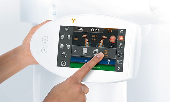
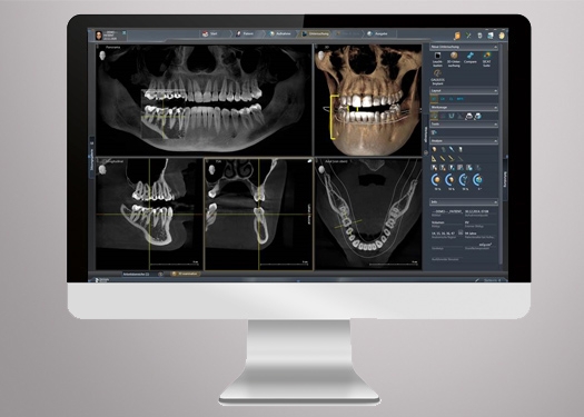
The 3D Advantage at a Glance

  • Improvement in image quality
  • Easy positioning to expose better quality images
  • Confidence that all the clinical details are seen
  • No more retakes that expose patients to additional radiation
  • Advanced filtering enhancements & optimizations
  • Support for an accurate diagnosis

Learn more >

Orthophos 3D X-Ray Machines

The level of data delivered from a 3D image is a revolutionary force in dental diagnostics. The ability to visualize overlaid teeth, unexpected canals, and hidden roots elevates the accuracy and impact of a patient’s diagnosis. Enhancing understanding and confidence positively influences patient acceptance and treatment success.

Efficient Diagnostics + Exceptional Image Quality

Always following the ALARA principle, our 3D solutions make sure that you can count on the image quality you need for the given indication at the lowest possible dose.

Reproducible + Easy Handling

Our innovative patient positioning systems and intuitive control panel reduce the learning curve previously required for optimal imaging results. Integrate any of our units into your practice with confidence, immediately increasing the efficiency and accuracy of your diagnostic processes.

Proven Reliability + Quality

With more than 100,000 units sold worldwide, we can say one thing for sure, our Orthophos units stand for best-in-class reliability. High German quality standards, consistent product reliability, and ease of operation provide owners with a dynamic tool to add into their practice. Reliable after-sales support and topical product training provide additional peace of mind.

Broader Practice Offering + Patient Acceptance

Our smart treatment solutions are perfect companions for our x-ray units, giving you new possibilities for your practice offering and seamlessly adapting to future growth. Provide the visual evidence your patients need to become advocates for their own oral health.

Orthophos S

The Orthophos S is a high-quality X-ray unit with a comprehensive range of services optimized for everyday tasks. Orthophos S 3D is a reliable partner with automatic patient positioning with its patented occlusal bite block. The CsI Plus sensor with autofocus function produces sharp images even in anatomically difficult cases. For use in orthodontics, the Orthophos S is also available with an optional ceph arm that can be retrofitted at any time.

Orthophos SL

The premium SL 3D unit is just right for practices with a keen understanding of the latest technologies. The integrated Direct Conversion Sensor (DCS) and Sharp Layer technology redefines the standard of panoramic imaging, providing exceptional performance even in difficult cases. Orthophos SL guarantees maximum ease of use through automatic positioning, intuitive operation with the EasyPad, and an individually adjustable ambient light for an exclusive look and feel. For bitewing, sinus or ceph images, left or right ceph arms are optional and can be retrofitted at any time. 

Orthophos 3D Imaging

Custom 3D Imaging

A significant advantage of Orthophos 3D technology is the ability to achieve precise images even when addressing demanding clinical situations, delivering precision and versatility. With Orthophos 3D X-ray Units, each volume can be adjusted according to three different modes to adapt to a patient’s specific situation: High-resolution volumes (HD), Minimal dose setting (Low Dose), or Standard Definition (SD).

Low Dose Mode

The Low Dose Mode, with a dedicated filter, delivers CBCT imaging in the dose range of a 2D image. In a single scan see clear, detailed, three dimensional images of teeth, soft tissues, nerve pathways, and bone at a greatly reduced dose. Cone Beam imaging has become increasingly important in treatment planning and diagnosis in implant dentistry, ENT, orthopedics, and interventional radiology (IR).

High-Definition Mode

When you require better visibility of fine structures for treatment planning and implementation, Orthophos offers you High Definition Mode (HD). The 3D X-ray system records 800 individual images during one rotation and merges the data into a low-noise 3D volume with high resolution of up to 80 μm. This mode guarantees a faster and safer diagnosis within the recorded volume.

Metal Artifact Reduction Software (MARS)

Metal artifacts are a challenge in 3D imaging. Radiopaque objects create shadowing and streaking effects during the three-dimensional reconstruction and interfere with findings. MARS automatically reduces metal artifacts and keeps anatomically relevant structures as clear as possible, facilitating a more comprehensive diagnosis.

Sidexis 4 Software

The modern and highly intuitive imaging software – Sidexis 4 – supports clear diagnoses. With its award-winning user interface, it promotes an accessible workspace and easy navigation, saving valuable practice time. The versatile platform also creates a basis for optimal patient communication, increasing patient comfort and understanding.

Sidexis 4 compares two CBCT images or up to four 2D images simultaneously. For example, you can navigate through both volumes at the same time, obtain cross-comparisons, and provide clear patient communication and case presentation. This saves you valuable time and enhances confidence in the diagnosis and treatment.

Intraoral Scanners

When looking for a digital impression solution, Primescan sets a new standard in dental scanning technology. Primescan scans at an incredible data density, delivering complete 3D structures of everything in its field of view – right from the very first scan.

Digital Accuracy

Dynamic depth scan technology enables perfect sharpness and outstanding precision, even at a measuring depth up to 20 mm.

Exceptional Hygiene

With three different sleeve concepts, it has never been so easy and hygienic to take digital impressions. Delivering the same level of performance, reprocessing options include disinfection, autoclavable, or disposable. A fully closed scanning window prevents liquid infiltration during the scanning process.

Usability

The new Acquisition Center is a workstation designed for modern dentistry and with the dentist in mind. It comes with a touchpad and a 16:9 wide-format movable touchscreen, offering you a highly intuitive and ergonomic work platform.

Efficiency

Primescan intraoral scanner allows you to start scanning right away. It offers continuously self-heating for fog-free scanning. Thanks to the increased field of view, you’ll be able to visualize larger areas with less sweeps and with immediate precision. The excellent scan results are instantly displayed on the touchscreen of the Acquisition Center for patient consultations.

Primescan

The innovative Smart Pixel Sensor processes more than 1,000,000 3D points per second that produces photorealistic and highly accurate data. Its dynamic depth scan technology enables perfect sharpness and outstanding precision, even at a measuring depth up to 20 mm – an absolute advantage for deeper-lying indications.

Immediate analysis allows for quick identification of required touchups, as well as material choice and viable preparations. A secure connection seamlessly delivers the high-resolution 3D impression directly to a preferred lab or specialist. Practitioners can consult directly with a dental technician even during treatment or initial consultation.