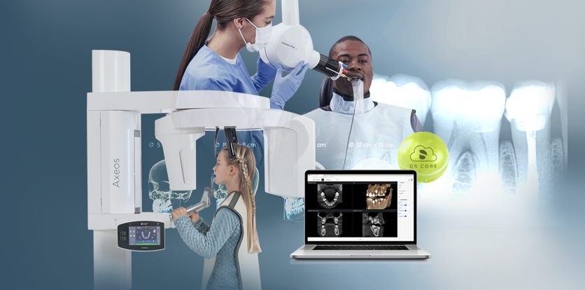
Get started in digital imaging
The sound 2D X-ray unit providing a smooth entrance into the world of digital imaging. Thanks to CsI sensor technology and its easy-to-use interface, you are ensured reliable diagnostics every time. The cephalometric option also makes the Orthophos E a reliable partner for orthodontics. Enrich your practice with a wide range of services that are only possible with digital imaging.
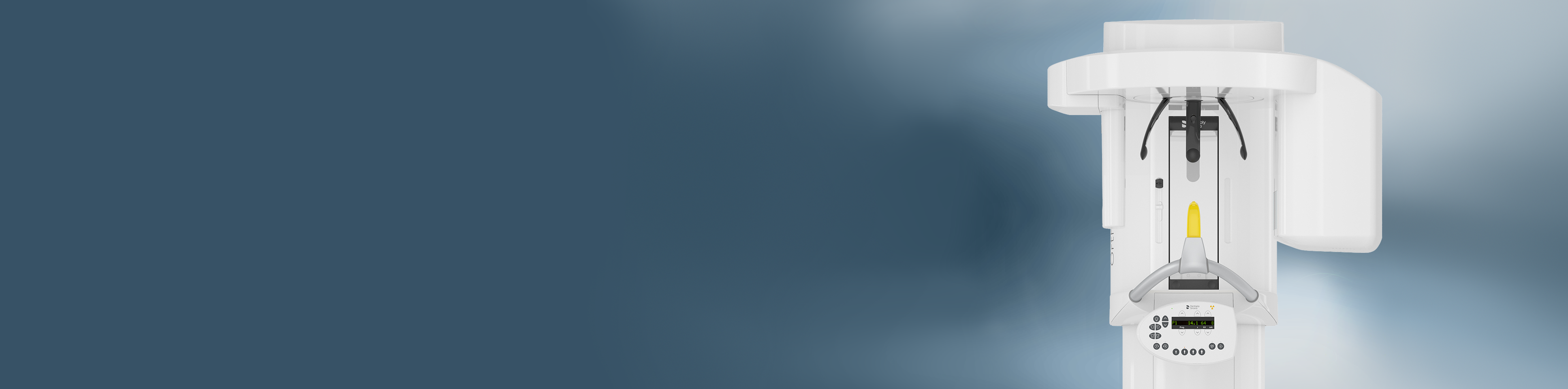
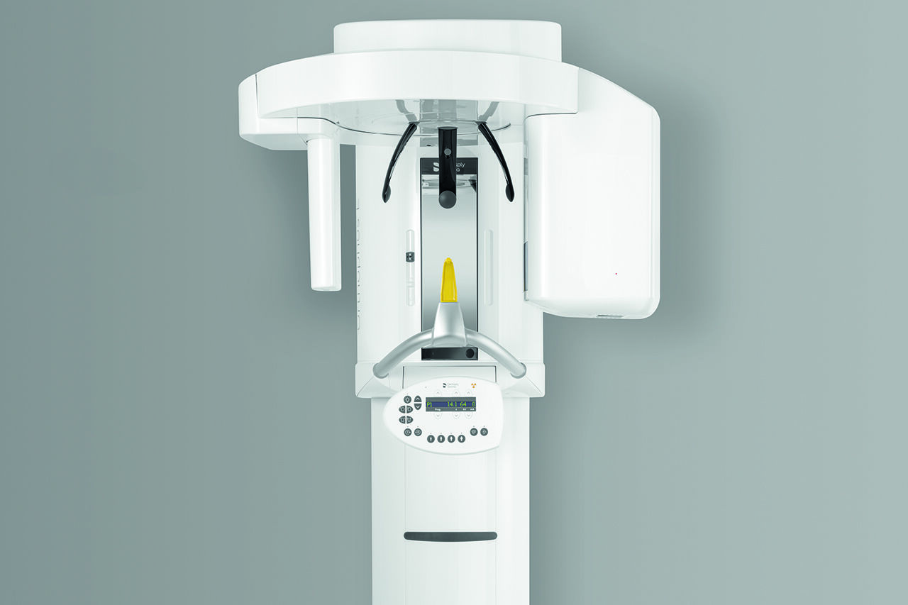
Orthophos E from Dentsply Sirona
Benefits
Why Choose the Orthophos E?


Reliable diagnosis
Thanks to the 2D CsI sensor and reliable image quality
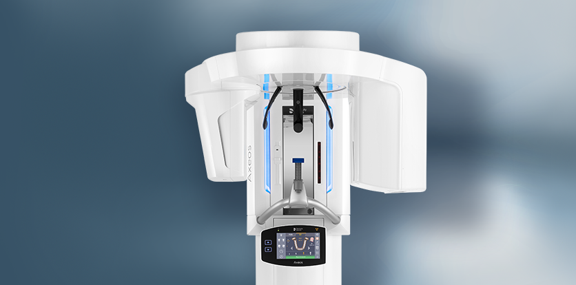
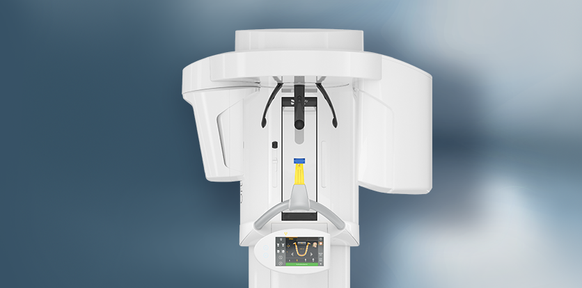
Safe and proven patient positioning
With motorized temple and forehead support, automatic temple width measurement, light localizers and built-in handles for positioning stablization
Essential 2D programs
For basic diagnostics in 2D
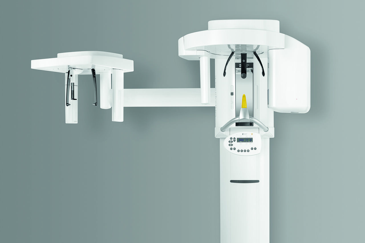
Ceph images
For optimal diagnostic support in orthodontics a left ceph arm can be ordered or retrofitted at any time
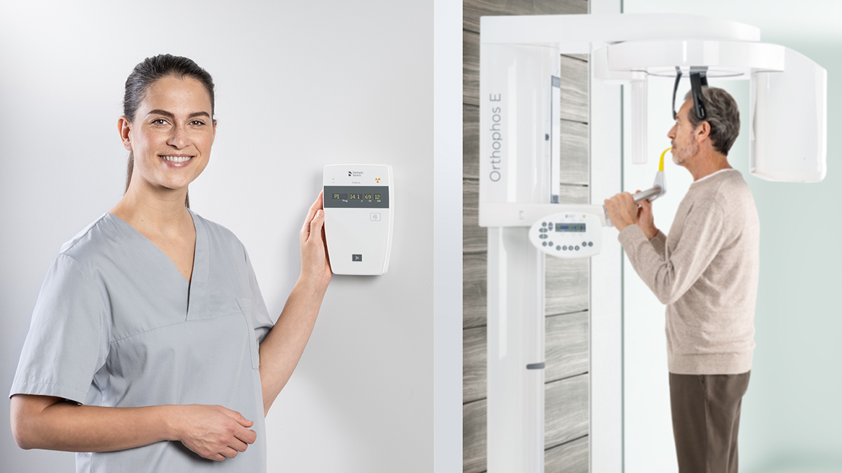
Easy and intuitive operation
With the MultiPad control panel
Proven Patient Positioning
Orthophos E has two integrated light localizers to facilitate ideal positioning in the sharp layer. The motorized forehead and temple supports gently fix the patient’s head in place and provide additional support to prevent motion blurring.
Gallery of Sample 2D Panoramic Images
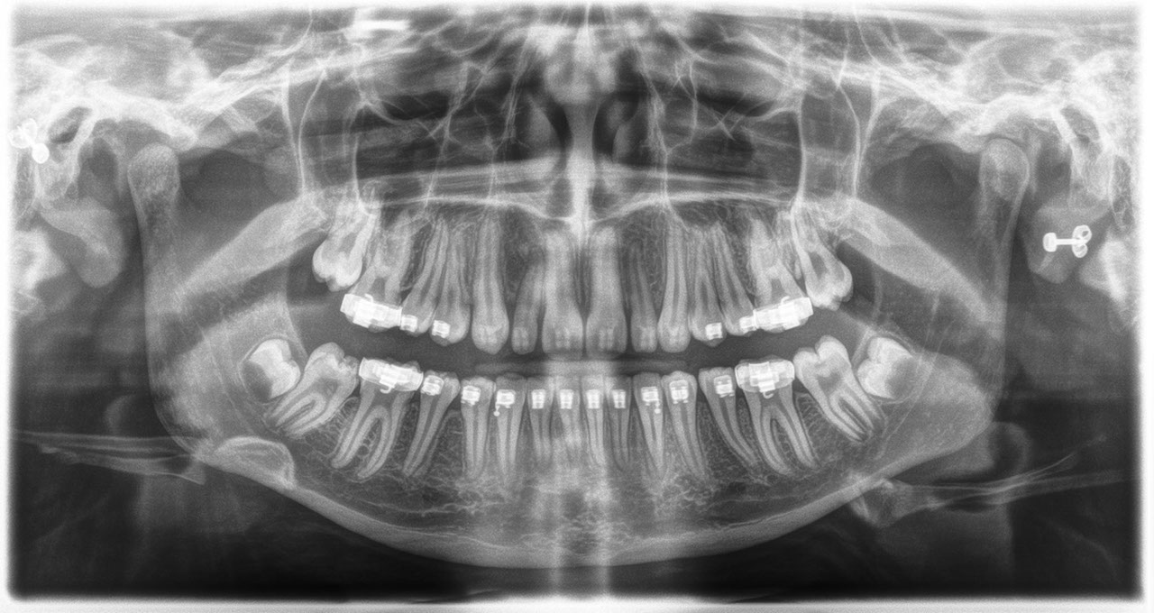 Adolescent, full dentition with orthodontic braces. Agenesis of teeth 18 and 28 with the presence of odontoblasts in 38 and 48. The incomplete root growth in 37 and 47 as well as the root development of the two wisdom teeth are evidence of the patient's young age. Maxillary sinuses and surrounding anatomical structures not radiographically apparent
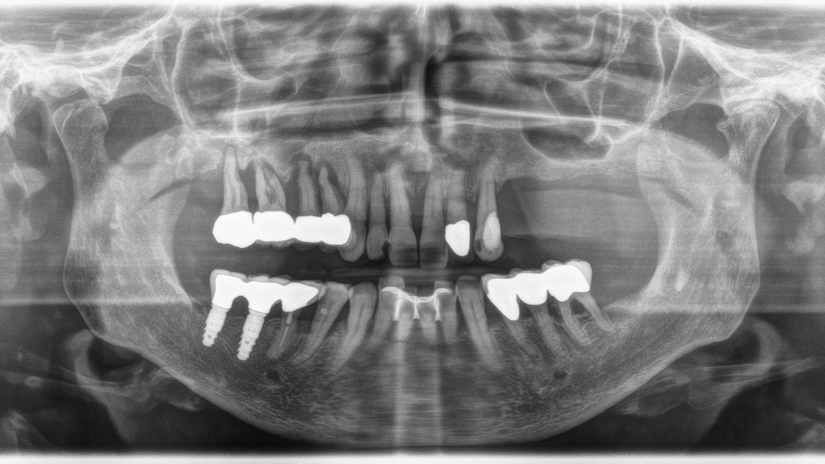
 Prosthetic and prophylactic treatment of dentition. Hypodense clearly limited changes to the hard tooth substance in teeth 12, 11 and 22 (presumably non-x-ray-opaque composite fillings here). Hyperdense filling material in the roots of teeth 11, 36 and 35 without an indication for periapical lucency. Status post apicoectomy on tooth 36 with retrograde obturation of the mesial and distal roots. Good osseous union of the apical bone tissue, no indication of recurrent periapical osteolysis
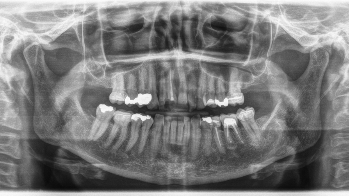
 Dentition badly damaged by periodontitis, with generalized horizontal bone resorption and vertical cavities with teeth 17 – 14 requiring extraction. Hypodense, homogenous and clearly delimited changes to the hard tooth substance on 12 mesial, 11 distal and 23 mesial with suspected caries or a differential diagnosis of non-x-ray-opaque composite fillings. Differential diagnosis: Caries or non-x-ray opaque composite fillings. PFM bridge at teeth 36 – 34 due to furcation on tooth 36 and calculus deposits mesially. Splinting of teeth 32-42 from past periodontal history. Hyperdense filling material in tooth 45 without an indication for periapical lucency. Implant-based treatment in the region of teeth 46 and 47 with radiographically inconspicuous PFM hybrid bridge 45 – 47
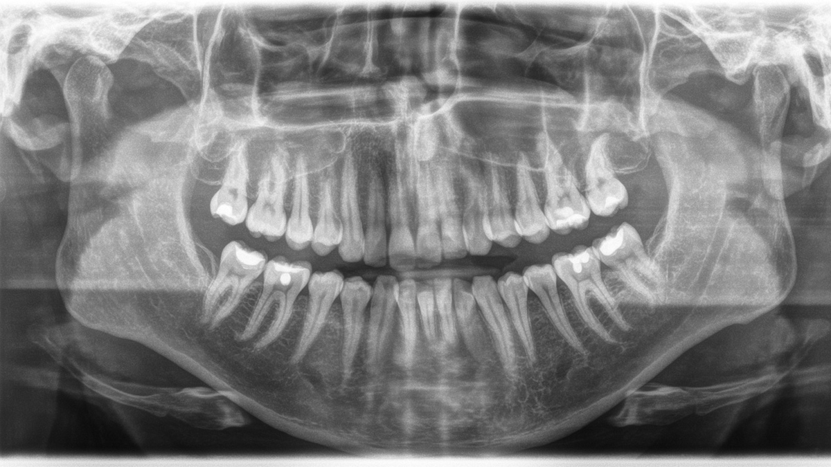 Prophylactically treated full dentition. Wisdom teeth are not present. Large maxillary sinuses are visually inconspicuous. The adjacent anatomical structures are also radiographically inconspicuous.
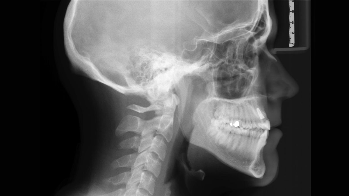
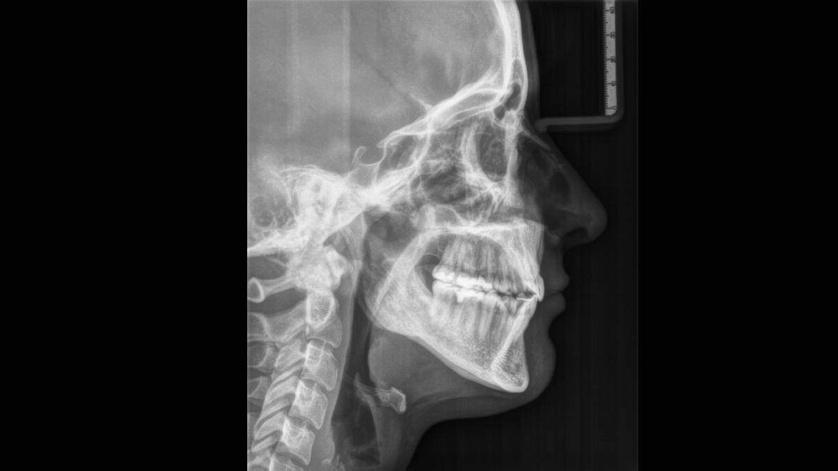
Sample Gallery of Cephalometric Images
FAQs
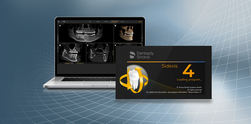
For more details please see Sidexis 4 system requirements.
Dentsply Sirona X-ray units work exclusively with Sidexis 4. Nevertheless data migration from Sidexis XG to Sidexis 4 is very easy. Sidexis 4 allows for the full digital experience with the latest tools