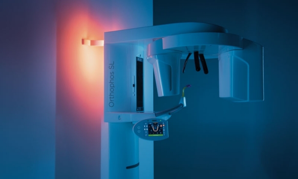
Ambient Light
With a selection of over 30 colors, the Ambient Light of the Orthophos SL 2D creates a pleasant atmosphere for your patient and blends in perfectly with your modern practice look.
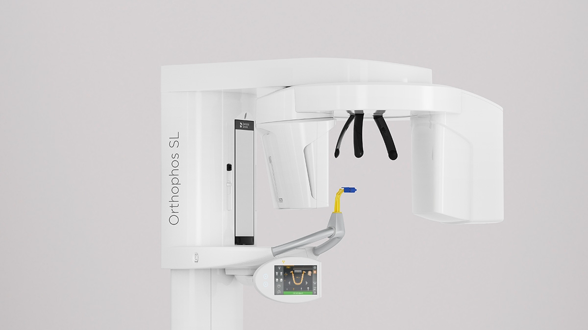
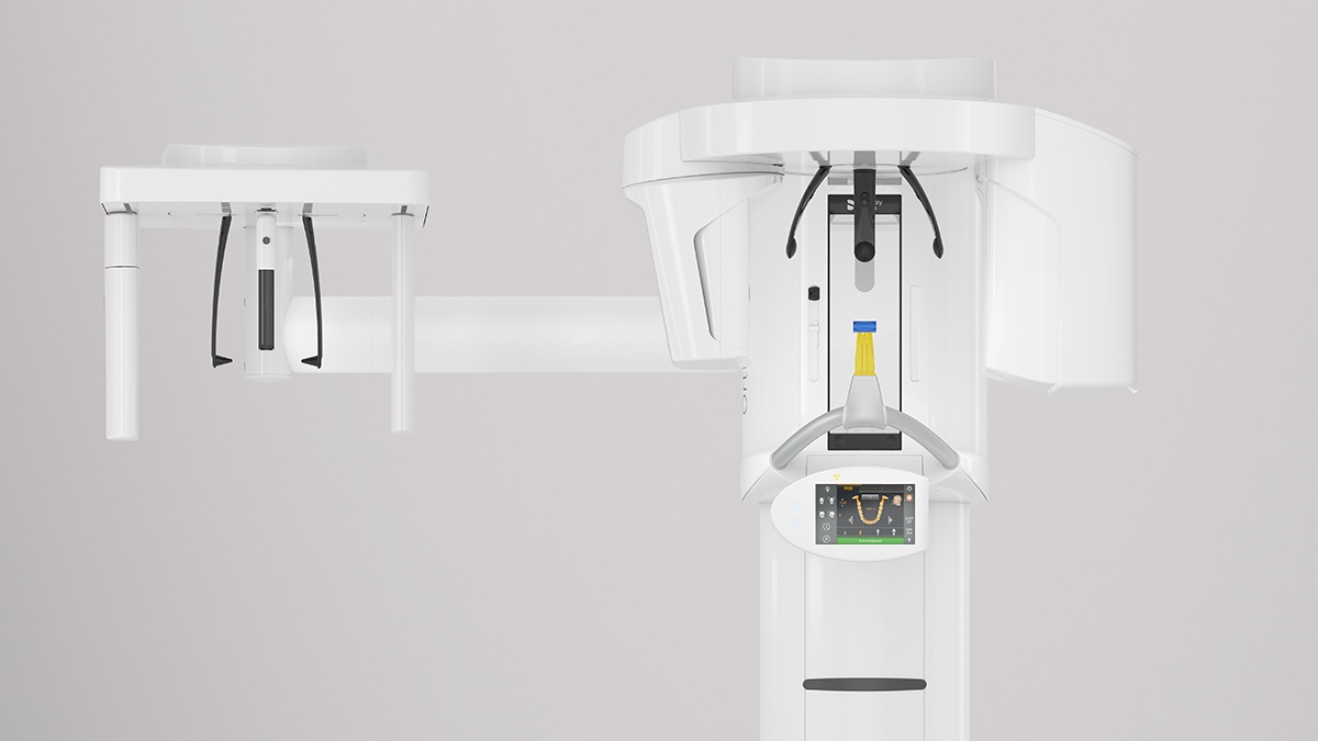
The top-quality 2D high-end device for practices with a keen understanding of the latest technologies and for those who simply want more. The integrated Direct Conversion Sensor (DCS) completely redefines the standard of panoramic imaging – delivering unique sharpness. The namesake, the Sharp Layer technology, provides autofocused panoramic images, even in difficult cases. The Orthophos SL guarantees maximum ease of use through automatic positioning, intuitive operation with the EasyPad and offers an individually adjustable ambient light for an exclusive look and feel.
With the Orthophos SL 2D, you’re choosing the best 2D image quality – and you’ll be ready to move to 3D at any time.
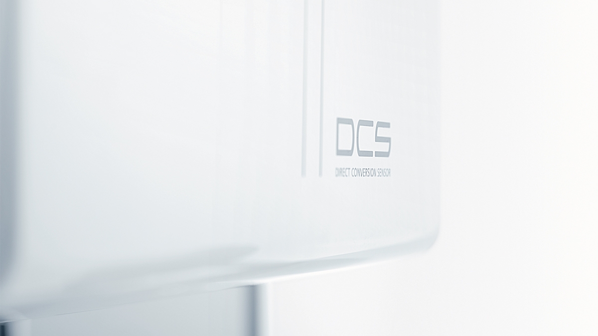
The unique DCS sensor for the best image quality
The Sharp Layer technology offers the possibility of displaying in reliable sharpness on different levels
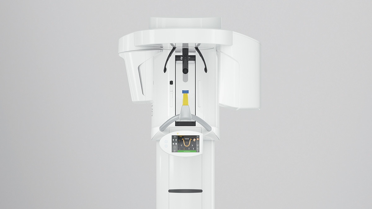
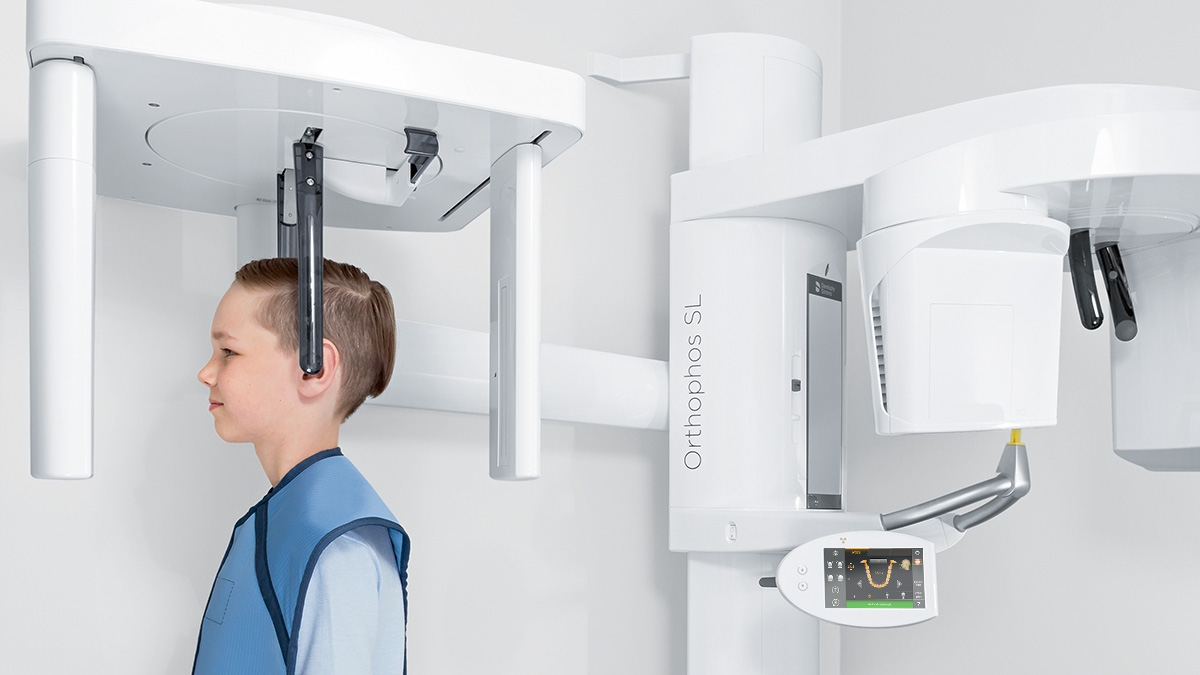
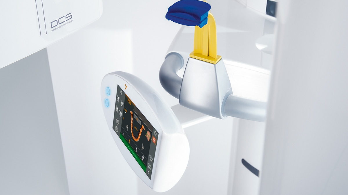
The auto positioning with the occlusal bite block and the intuitive EasyPad provide stability
For bitewing, sinus or Ceph images, left or right ceph arms are optional and can be retrofitted at any time
With motorised temple and forehead support, automatic temple width measurement, light localisers and sturdy handles
The ambient light is available in over 30 colors
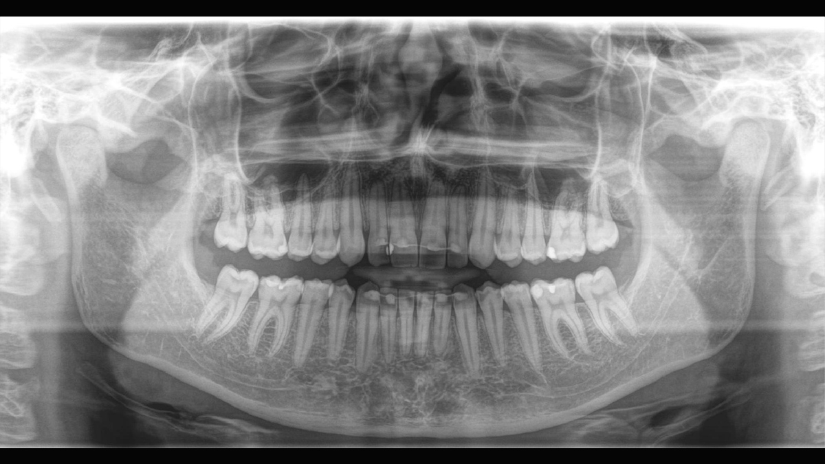
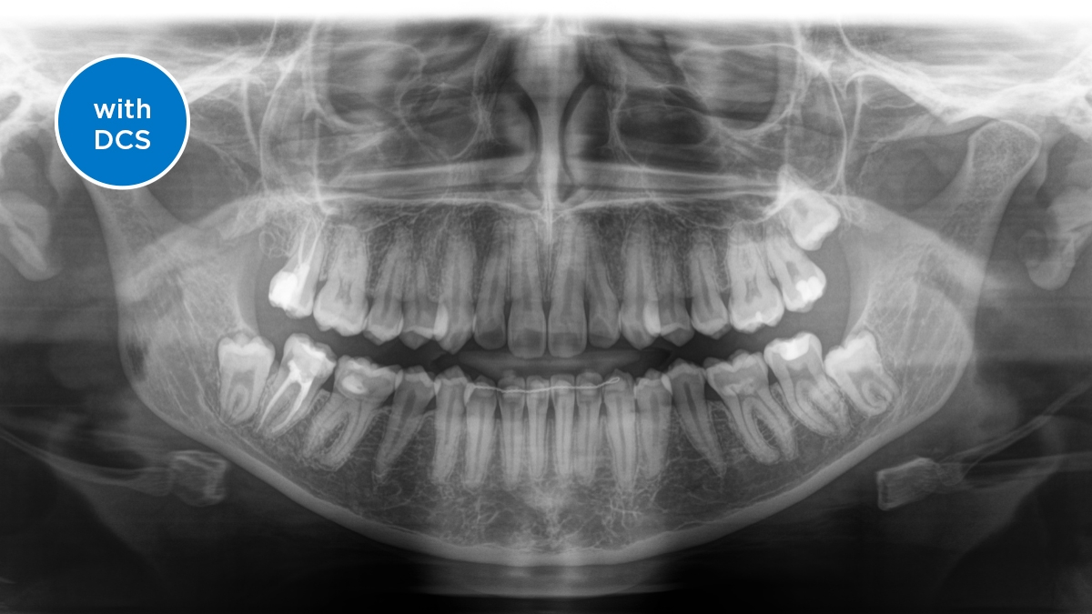
The Direct Conversion Sensor (DCS) ensures the optimal yield of X-ray radiation. The images are very precise – and it is possible to reduce the radiation dose.
Orthophos SL - Program P1
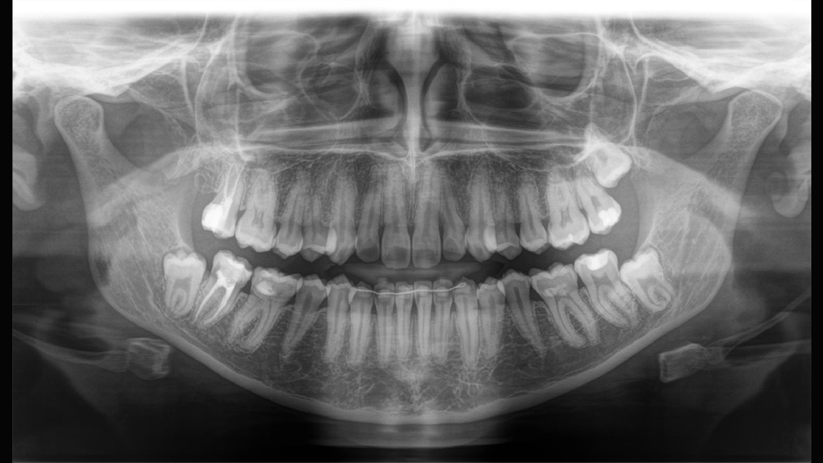
Orthophos SL - Program P1
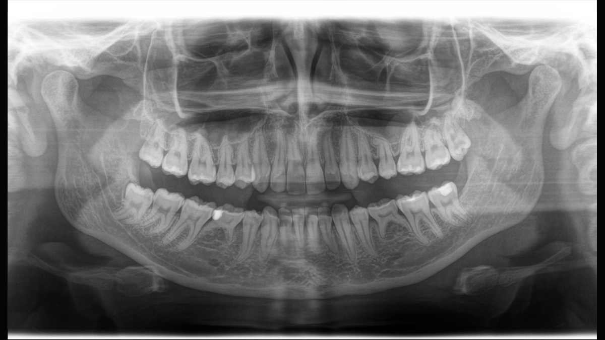
Orthophos SL - Program P2
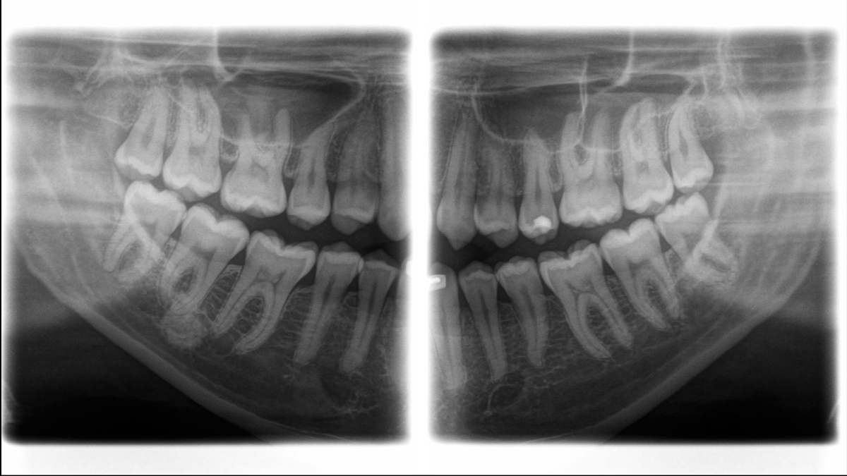
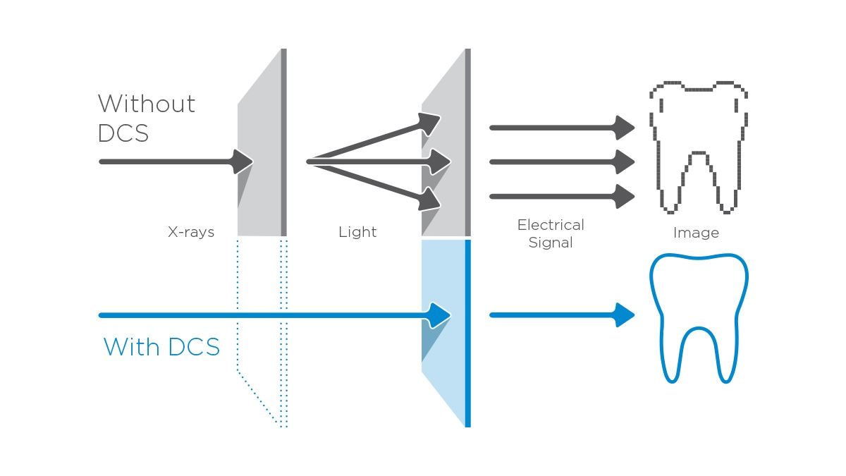
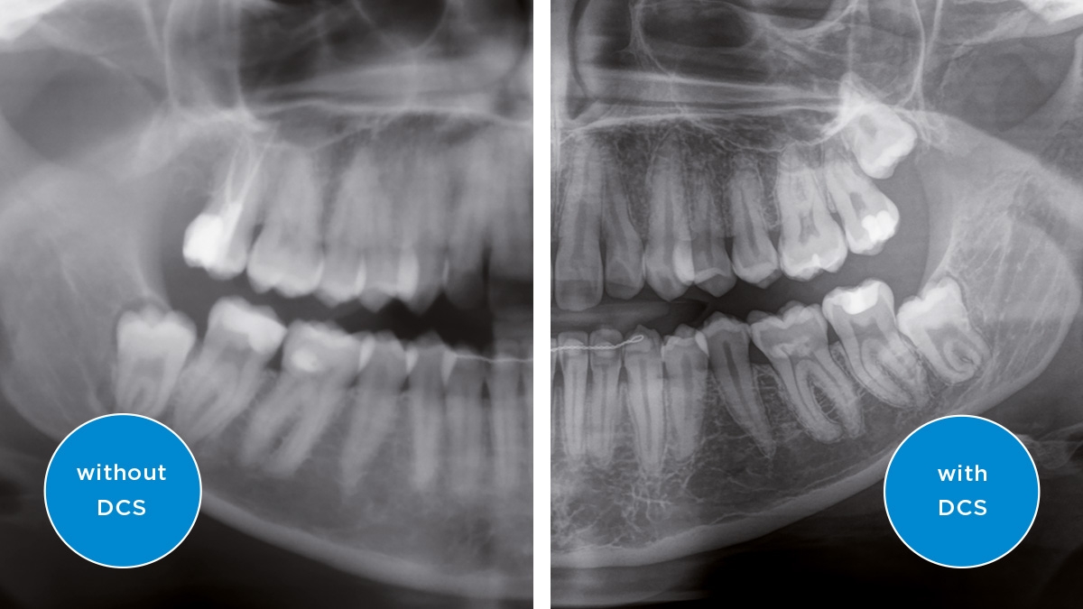
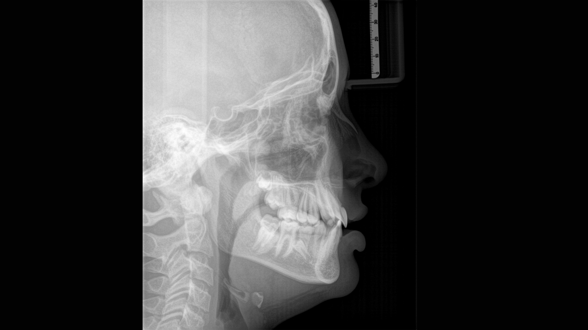
The Direct Conversion Sensor (DCS) has redefined the standard of panoramic imaging. X-rays are converted directly into electrical signals – unlike conventional systems, there are no signal losses due to light conversion. This means an improved image information output for you. The result is images with a uniquely high level of sharpness – even at an extremely low dose.
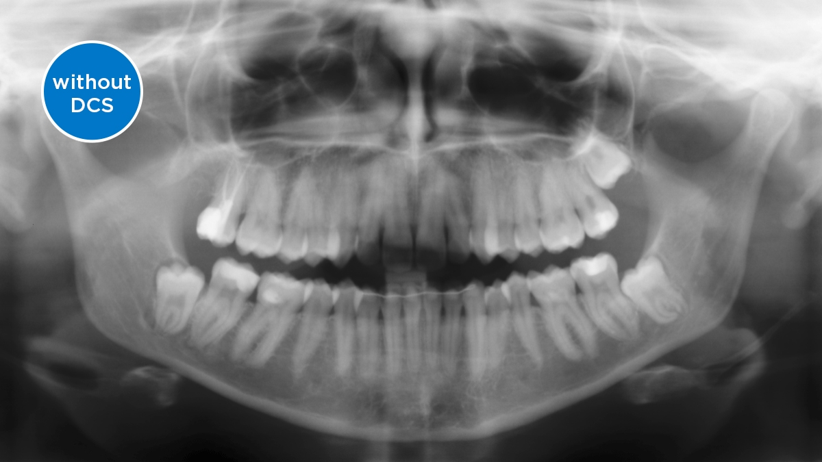
The image shows X-ray images with and without DCS technology. Move the control over the image and see for yourself the difference the DCS sensor makes to the image quality.
With a selection of over 30 colors, the Ambient Light of the Orthophos SL 2D creates a pleasant atmosphere for your patient and blends in perfectly with your modern practice look.
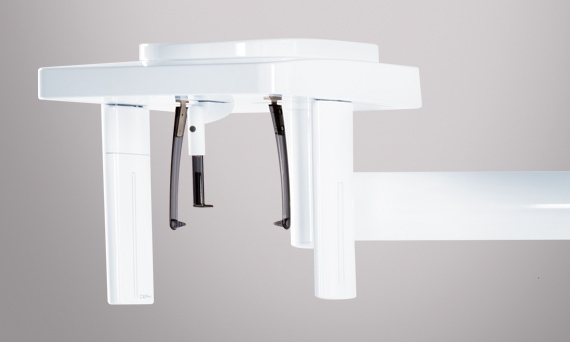
Orthophos SL 2D offers the capability to install a cephalometric arm at any time. In addition, to be sure it fits in your X-ray room; the arm can be mounted on either the right side or left side of the unit.
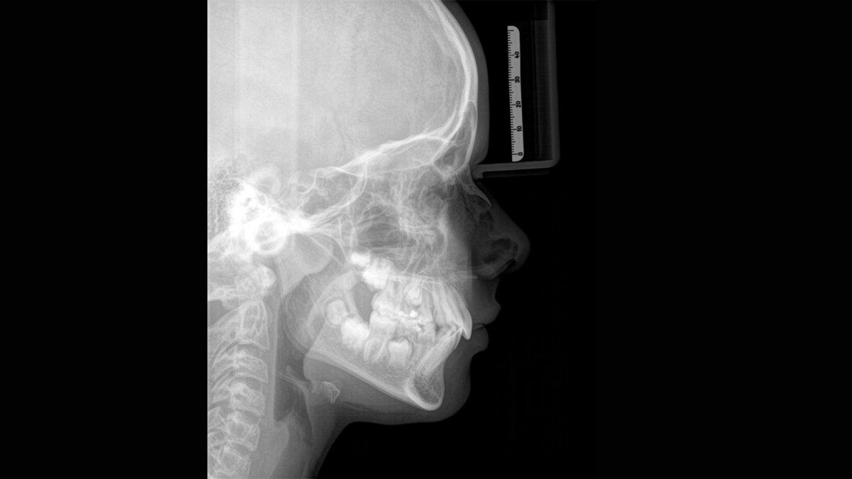
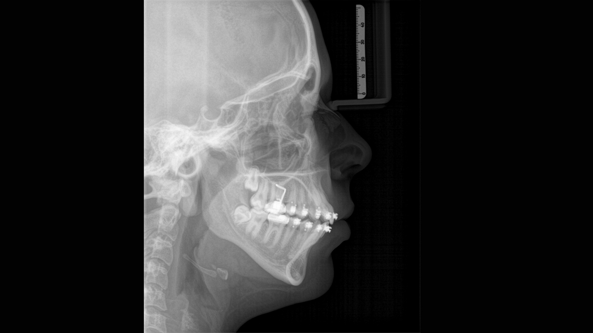
Using a dedicated sensor, you get lateral and symmetrical p.a. or a.p. as well as carpus images. In cases of displaced teeth, you can also fall back on the benefits of 3D x-rays to determine their exact location.
| Programme | Orthophos SL 2D | Orthophos S 2D | Orthophos E |
|---|---|---|---|
| Standard panorama image |
P1, P2, P10 |
P1, P2, P10 |
P1, P10 |
| Image detail left side or right side |
P1, P1A, P1C P2, P2A, P2CP10, P10A, P10C BW1 |
P1, P1A, P1C P2, P2A, P2CP10, P10A, P10C BW1 |
P1L, P1R |
| Image detail individual quadrants |
P1, P1A, P1C P2, P2A, P2CP10, P10A, P10C |
P1, P1A, P1C P2, P2A, P2CP10, P10A, P10C |
- |
| Image detail upper or lower jaw |
P1, P1A, P1C P2, P2A, P2CP10, P10A, P10C, P12 |
P1, P1A, P1C P2, P2A, P2CP10, P10A, P10C, P12 |
- |
| Constant magnification |
P1C, P2C, P10C |
P1C, P2C, P10C |
P1C |
| Artifact-reduced |
P1A, P2A, P10A |
P1A, P2A, P10A |
P1A |
| Thick layer front |
P12 |
P12 |
P12 |
| Sinusoidal images |
S1, S3 |
S1, S3 |
S1 |
| Multislice in posterior tooth |
- | - | MS1 |
| Mandibular joint |
TM1.1, TM1.2, TM3 |
TM1.1, TM1.2, TM3 |
TM1.1, TM1.2 |
| Bitewing image |
BW1, BW2 |
BW1, BW2 |
BW1 |
| Ceph (optional) |
C1, C2, C3, C3F, C4 |
C1, C2, C3, C3F, C4 |
C1, C2, C3, C3F, C4 |
The requirements actually follow those for the image processing software, Sidexis 4. For more details please see Sidexis 4 System Requirements.
Yes, the Orthophos SL works exclusively with Sidexis 4. Nevertheless data migration from Sidexis XG to Sidexis 4 is very easy.
Yes, it is possible. To proceed, the serial number of your unit will requested. Thus, a visit to your practice by a local Service Technician will need to be scheduled.
Find out more about our 2D and 2D/3D hybrid product range and download the extraoral X-ray family brochure here.
Whether hardware or software, a variety of solutions are waiting for you in extraoral x-rays.

Find out more about the imaging systems from Dentsply Sirona and request information on intraoral imaging, software or 2D and 3D imaging technology.