
Llámenos o proporcione sus datos de contacto a continuación, y un representante de Dentsply Sirona se pondrá en contacto pronto.


Contáctenos
Lleve a su clínica al siguiente nivel.
Descubra más formas de aportar innovación a su clínica.
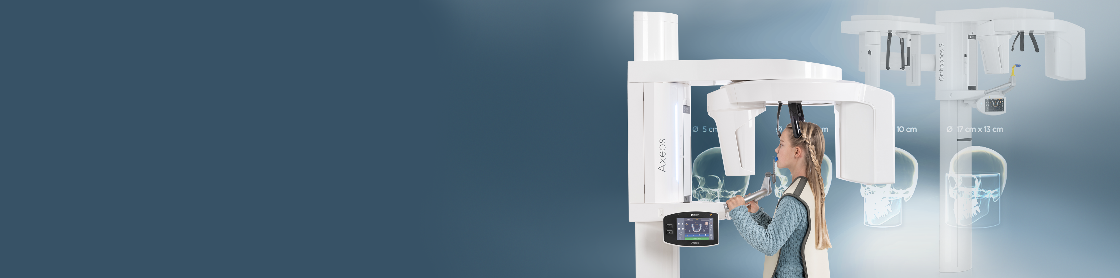
Diagnóstico de boca completa potenciado por dispositivos prémium
Mejore la salud bucal de sus pacientes al tener la capacidad de diagnosticar y planificar el tratamiento para casi todas las etiologías en la región dentomaxilofacial. Las soluciones en radiología de DS apoyan las necesidades del paciente, desde la captura de las articulaciones temporomandibulares hasta la visualización de los detalles más pequeños utilizando radiografías de endodoncia de alta resolución de hasta 80 µm.
Cuando se combinan con las capacidades de DS Core y el robusto software de radiología, los clientes de Dentsply Sirona tienen la confianza necesaria para brindar una mejor atención al paciente.
NUESTROS PRODUCTOS
Descubra nuestros productos de imágenes radiológicas extraorales


BENEFICIOS
¿Por qué elegir equipos de radiología extraoral DS?

Mejore sus capacidades de diagnóstico.
Obtenga una posición del paciente de alta calidad.
- Reduzca la necesidad de repetición de tomas gracias a nuestras características de posicionamiento.
- Radiografía de alta calidad incluso con artefactos gracias a MARS, nuestro software de reducción de artefactos metálicos
- Resalte pequeños detalles debido a nuestra resolución de hasta 80 μm.
- Reduzca el riesgo de artefactos por movimiento gracias a nuestras características de posicionamiento del paciente.

Empodere el crecimiento de la clínica
Ofrezca una amplia gama de tratamientos y tareas a su equipo para enfocarse en sus pacientes.
- Cubra las necesidades de radiografías de sus pacientes internamente gracias a los campos de visión en expansión de nuestras unidades.
- Disfrute de la tranquilidad de saber que su personal puede capturar radiografías de alta calidad con nuestros dispositivos avanzados de posicionamiento del paciente.
- Aumente la seguridad del paciente utilizando radiografías de baja dosis para pacientes sensibles y cuando la alta definición no es crítica para un diagnóstico preciso.
- Prospere en su planificación de tratamiento de endodoncia gracias a nuestro modo 3D de alta definición de hasta 80 μm.
TESTIMONIO
Comentarios de los profesionales dentales
"Gracias al Posicionador automático, los tiempos de espera se reducen".

Dr. Bernhild Elke Stamnitz
GP y especialista en cirugía dental y estética
CONTENIDO RELACIONADO
Haga más con su unidad de radiología extraoral DS con DS Core.

DS Core
Acceda al universo digital de soluciones de Dentsply Sirona través de DS Core
La primera plataforma interconectada y digitalmente avanzada de Dentsply Sirona está diseñada para brindarle un apoyo completo durante toda la historia clínica del paciente, desde el diagnóstico hasta la atención o tratamiento final. Esto le permite utilizar flujos de trabajo continuos, fomentar la colaboración con socios y mantenerse al tanto de los últimos desarrollos en odontología digital. Benefíciese de las funcionalidades en la nube de DS Core y de la integración directa con su clínica de inmediato.
Descubra más soluciones en radiología

Imagen radiológica intraoral
Evaluación confiable potenciada por la precisión

Software de imágenes radiológicas
Cada tratamiento comienza con un diagnóstico preciso
