image-comparison grip-lines arrow-top-right zip arrow-right video minus arrow-back text shop twitter file home quick-order arrow-left youtube search arrow-down check all filter site_references image Slice 1 zoom 🎬 icons / _ icon size / _ download-queue Created with Sketch. chevron-down arrow-right picture pdf podcast exe plus hotline close xxx-1 burger clipboard linked-in ppt Artboard 🎬 icons / _ icon size / _ download-queue_add Created with Sketch. Layer 1 Created by potrace 1.15, written by Peter Selinger 2001-2017 My Account press_releases Created with Sketch. PRESS ds-instruments ds-preventive ds-implants ds-endodontics Cart presentation Created with Sketch. DS-Preventive-Icon-Grey video Created with Sketch. other Created with Sketch. text_piece Created with Sketch. promotion Created with Sketch. Spare Parts ds-imaging-systems case_study Created with Sketch. ds-treatment-centers broschure Created with Sketch. ds-prosthetics ds-restorative video Created with Sketch. flyer Created with Sketch. whitepaper Created with Sketch. article Created with Sketch. image Created with Sketch. Service ds-cad-cam Artboard Quick Order Dentist Chair study Created with Sketch. ds-orthodontics ds-implants ds-endodontics ds-cerec digital-scan grinder ds-diagnosis END-Website-Icons-Shaping-Cleaning-Obturation step_1 analyse ds-abutment-placement more-quality initiative scan more-quality ds-polishing digital ds-irrigation ds-treatment-centers Test treatment ds-restorative ds-implant-planning realistic construct book ds-planning-impression END-Website-Icons-Shaping-Cleaning-Obturation END-Website-Icons-Shaping-Cleaning-Obturation mirror anwender ds-preventive planning-2 ds-obturation ds-cad-cam 04 ds-implant-placement ds-retention-protection ds-anesthetics restauration form ds-inlab oven CORP-icon-grey-screen-tooth-cavity-pulpa robot plan place ds-orthodontics PDF-Formular_ganzneu PDF overlay ds-bone-regeneration ds-treatment-preparation vorher_nachher ds-maging-systems wifi END-Website-Icons-Shaping-Cleaning-Obturation omnicam PDF-Formular_neu_vonGünther treatment consulting planning comparison ds-set-up ds-prosthetics support step_3 ds-tooth-preparation ds-treatments ds-scaling step_2 ds-instruments software quote-english quote-french quote-text-german Created with Sketch. upload Created with Sketch. version Created with Sketch. ds-endodontics globe
Dentsply Sirona Norway
Norsk bokmål
Home Back

Xios XG Supreme

Excellent Images for Optimal Patient Care

Excellent image quality is just the beginning with the Xios XG Supreme intraoral sensor. With a number of enhancement options and the ability to easily adjust your images to support your individual preferences, you get more than just a sensor; you get an intraoral solution. Equipped with multiple sensor sizes, cable lengths, and connectivity options, you truly have a tool that adjusts to you, not the other way around. 

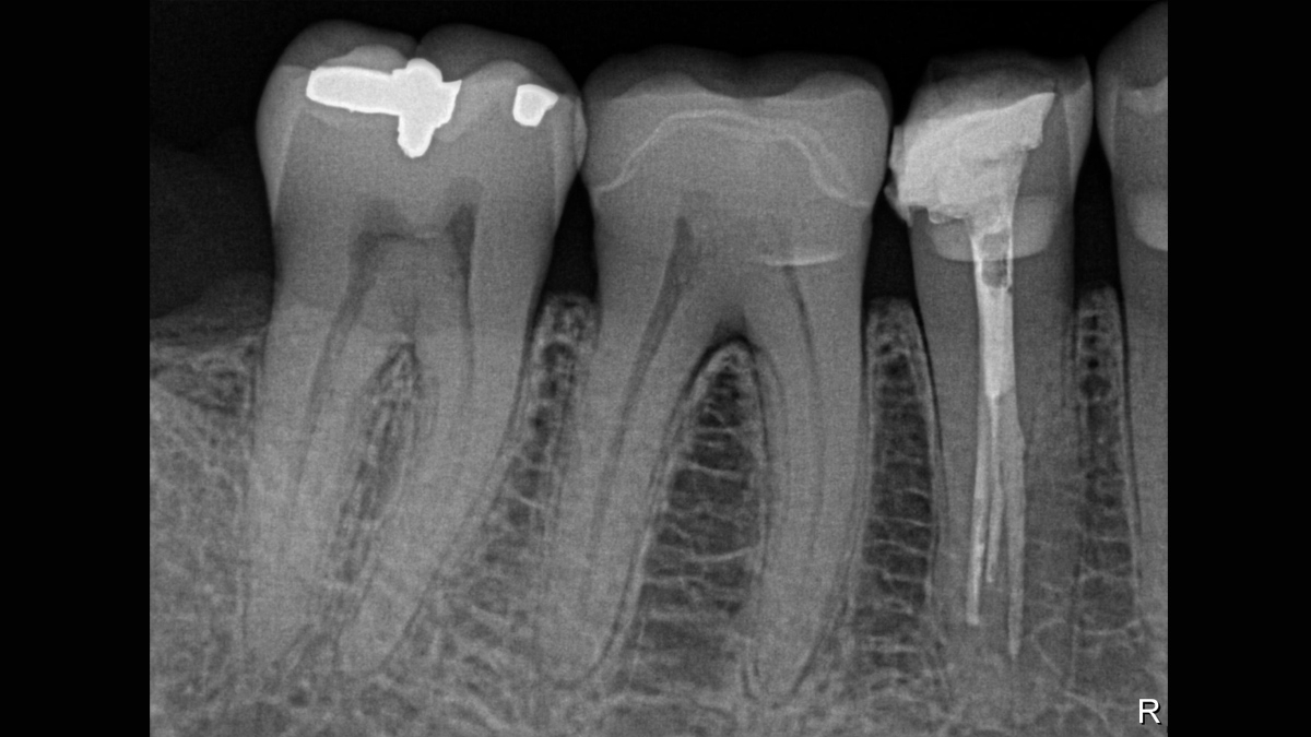
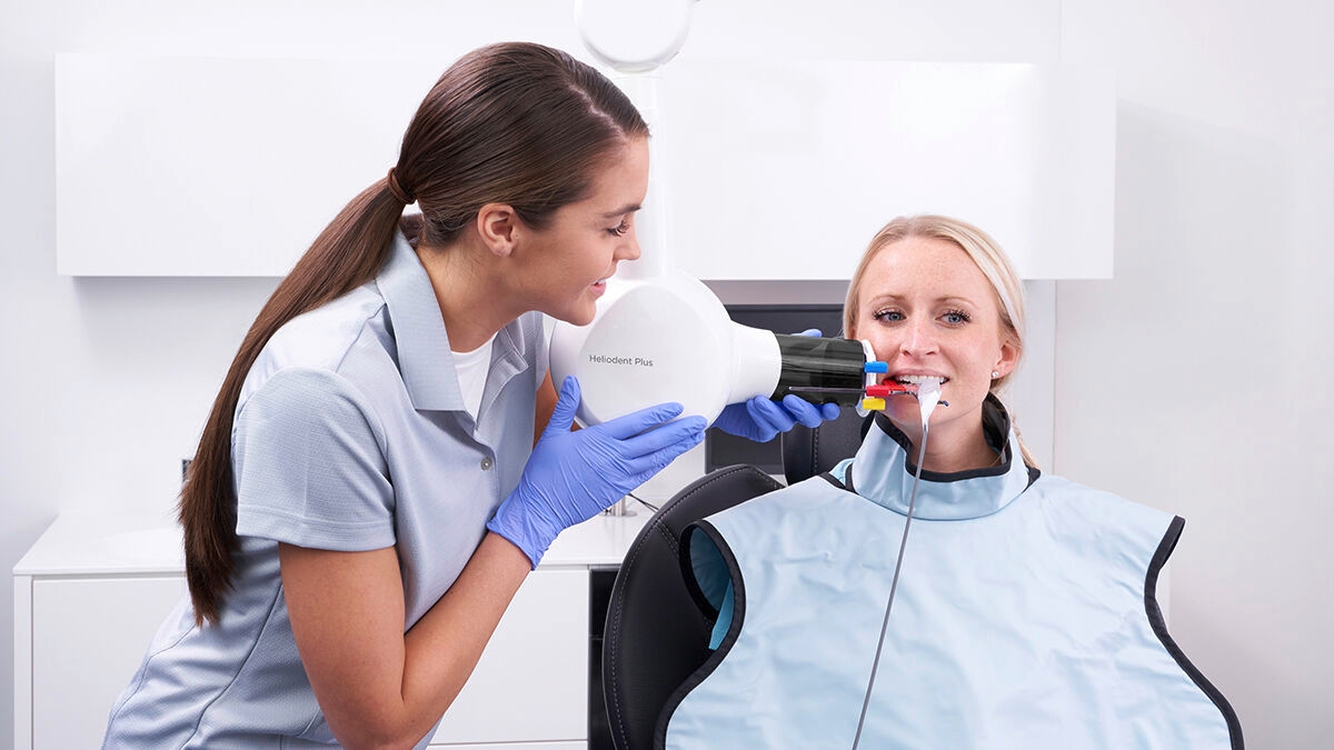
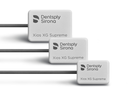
Xios XG Supreme from Dentsply Sirona

Size 0

  • Active area: 18 mm x 24 mm
  • Sensor dimensions: 23.6 mm x 32 mm x 7.5 mm
  • Ideal for pediatric images

Size 1

  • Active area: 20 mm x 30 mm
  • Sensor dimensions: 25.4 mm x 38.3 mm x 7.5 mm
  • Ideal for single tooth images for smaller adults, patients with a shallow palate and bitewings for larger children

Size 2

  • Active area: 25.6 mm x 36 mm
  • Sensor dimensions: 31.2 mm x 43 mm x 7.5 mm
  • Ideal for adult bitewings and single tooth images

Easy Cable Exchange

Ease cable is available in 3 lengths. If a cable gets damaged, you can change it completely in-house and within minutes. Replacing cables is as simple as loosening two screws, saving time and helping to minimize lost productivity while protecting your investment.

  • 90 cm
  • 180 cm
  • 270 cm

Benefits at a Glance

Your Xios XG Supreme intraoral sensor may quickly become your favorite tool to work with - a reliable and efficient addition to any practice. The benefits it provides are endless.

Excellent Image Quality

Xios XG Supreme provides a theoretical resolution of 33 lp/mm and a 15 μm pixel size delivering superb image quality without the additional wait time caused by a developing or scanning process.

Robust Design and Security of Investment

The Xios XG Supreme intraoral sensor has been engineered and tested to withstand years of regular daily use, ensuring a long product life cycle. The sensor's durable design results from the use of premium materials and high-standard production processes. Each sensor is equipped with a reinforced cable and an additional replacement cable.

Multiple Options to Fit Your Treatment Rooms

Xios XG Supreme has been designed for true modularity to support your specific way of working, offering 3 sensor sizes (0, 1, and 2) and 3 cables lengths (90 cm, 180 cm, 270 cm).

Chairside Imaging

By integrating your X-ray generator and sensors with your treatment center, you can truly leverage the concept of complete chairside dentistry.

Optimized USB Data Transfer

The new USB 3 interface allows for accelerated information read-out and optimized data transfer between your sensor and imaging software. Protected connection plugs provide additional durability in daily practice use.

Proven image sharpness

Excellent image quality is just the beginning with Xios XG Supreme intraoral sensors. The dynamic sharpening slider allows you to adjust your diagnostic settings in terms of sharpness. Additional slider options are available for modifications in brightness and contrast. After configuration, images are displayed in an efficient way to enhance your diagnostic capabilities, saving you valuable time. With just the click of your mouse, you get the right image for every treatment situation and ultimate clinical support.

I use the Xios XG Supreme sensor because of its mobility; I can now easily switch treatment rooms with it. The replaceable cable is also a great feature, as before I used to have issues with broken cables.

Dr. Antoine Hirardot, Le Mans, France


Working with Xios Supreme: Filtering enhancements

With Xios XG Supreme sensors you have the option of five different filtering options: General Dentistry, Endodontics, Periodontics, Restorative and Hygiene. These clinically specific views are designed to optimize your diagnosis, enhancing the image to suit your needs.

Paralleling Technique

The paralleling technique is now standard for intraoral X-ray diagnostics. Its diagnostic advantages are clinically proven. The paralleling technique requires holders that align the sensor at a right angle to the central beam and thus parallel to the tooth axis.

Dentsply Sirona's sensors and holder systems are designed to make this process as simple as possible to provide an exceptional experience for both the user and the patient. For a successful, diagnostically sound X-ray and comfort while capturing the image. 


Sensor Positioning

You will find all of our positioning videos on the Dentsply Sirona Youtube channel.

Paralleling Technique

The paralleling technique is now standard for intraoral X-ray diagnostics. Its diagnostic advantages are clinically proven. The paralleling technique requires holders that align the sensor at a right angle to the central beam and thus parallel to the tooth axis.

Dentsply Sirona's sensors and holder systems are designed to make this process as simple as possible to provide an exceptional experience for both the user and the patient. For a successful, diagnostically sound X-ray and comfort while capturing the image. 


FAQ

Bending imaging plates can distort images and jeopardize the reproducibility of the image quality - plate manufacturers specifically state that plates should not be bent. Intraoral sensors are not as susceptible to scratches or distortion of the image, offer quick X-ray capture and are overall much more durable, offering the ultimate intraoral imaging experience.

The requirements follow those set for the Sidexis 4 imaging software. For more details, please see Sidexis 4 System Requirements.

The Xios intraoral sensor cable technology is backed by years of leading experience and offers outstanding durability. The ability to exchange the cable helps to limit loss in productivity if unexpected damage occurs. For example, if a chair rolls over the cable or someone mistakenly shuts it in a drawer with force, there is the potential that the cable could get damaged. For these cases, it is good to be prepared and the exchangeable cable will ensure that you are fully supported and can continue working with minimal delay.

The USB interface comes equipped with a 2 m USB cable, and additional optional sensor cables go up to 2.7 m to suit every operatory configuration and allow for optimum movement and flexibility. 

Dentsply Sirona's intraoral holder systems allow for a hygienic, comfortable and accurate imaging experience and can be purchased through your local dealer.


Intraoral Family Brochure

Find out more about all of our Dentsply Sirona intraoral imaging solutions.

Sensor Care Guide

Optimize your infection prevention - protect your patients and your investment.

Expand Your Imaging

Whether hardware or software, a variety of imaging solutions are waiting for you.


Technical Data

Xios XG Supreme Sensor 
Size 0: Active surface18 mm x 24 mm
Size 0: Overall dimensions 23.6 mm x 32 mm  x 7.5 mm
Size 1: Active surface20 mm x 30 mm
Size 1: Overall dimensions 25.4 mm x 38.3 mm x 7.5 mm
Size 2: Active surface25.6 mm x 36 mm
Size 2: Overall dimensions 31.2 mm x 43 mm x 7.5 mm
Physical pixel size
15 μm
Theoretical resolution33.3 lp/mm
Measured resolution28 lp/mm
Sensor cable lengthUp to max. 2.7m
Xios XG Supreme USB Interface 
USP port
USB 2
Power supply
via USB port 


Technical Documentation

Xios XG USB Interface and Sensors Operating and Installation Instructions


Contact us for more information

Find out more about the imaging systems from Dentsply Sirona and request information on intraoral imaging, software or 2D and 3D imaging technology.