image-comparison grip-lines arrow-top-right zip arrow-right video minus arrow-back text shop twitter file home quick-order arrow-left youtube search arrow-down check all filter site_references image Slice 1 zoom 🎬 icons / _ icon size / _ download-queue Created with Sketch. chevron-down arrow-right picture pdf podcast exe plus hotline close xxx-1 burger clipboard linked-in ppt Artboard 🎬 icons / _ icon size / _ download-queue_add Created with Sketch. Layer 1 Created by potrace 1.15, written by Peter Selinger 2001-2017 My Account press_releases Created with Sketch. PRESS ds-instruments ds-preventive ds-implants ds-endodontics Cart presentation Created with Sketch. DS-Preventive-Icon-Grey video Created with Sketch. other Created with Sketch. text_piece Created with Sketch. promotion Created with Sketch. Spare Parts ds-imaging-systems case_study Created with Sketch. ds-treatment-centers broschure Created with Sketch. ds-prosthetics ds-restorative video Created with Sketch. flyer Created with Sketch. whitepaper Created with Sketch. article Created with Sketch. image Created with Sketch. Service ds-cad-cam Artboard Quick Order Dentist Chair study Created with Sketch. ds-orthodontics ds-implants ds-endodontics ds-cerec digital-scan grinder ds-diagnosis END-Website-Icons-Shaping-Cleaning-Obturation step_1 analyse ds-abutment-placement more-quality initiative scan more-quality ds-polishing digital ds-irrigation ds-treatment-centers Test treatment ds-restorative ds-implant-planning realistic construct book ds-planning-impression END-Website-Icons-Shaping-Cleaning-Obturation END-Website-Icons-Shaping-Cleaning-Obturation mirror anwender ds-preventive planning-2 ds-obturation ds-cad-cam 04 ds-implant-placement ds-retention-protection ds-anesthetics restauration form ds-inlab oven CORP-icon-grey-screen-tooth-cavity-pulpa robot plan place ds-orthodontics PDF-Formular_ganzneu PDF overlay ds-bone-regeneration ds-treatment-preparation vorher_nachher ds-maging-systems wifi END-Website-Icons-Shaping-Cleaning-Obturation omnicam PDF-Formular_neu_vonGünther treatment consulting planning comparison ds-set-up ds-prosthetics support step_3 ds-tooth-preparation ds-treatments ds-scaling step_2 ds-instruments software quote-english quote-french quote-text-german Created with Sketch. upload Created with Sketch. version Created with Sketch. ds-endodontics globe
Dentsply Sirona Poland
Polski
Home Back

Orthophos S 2D

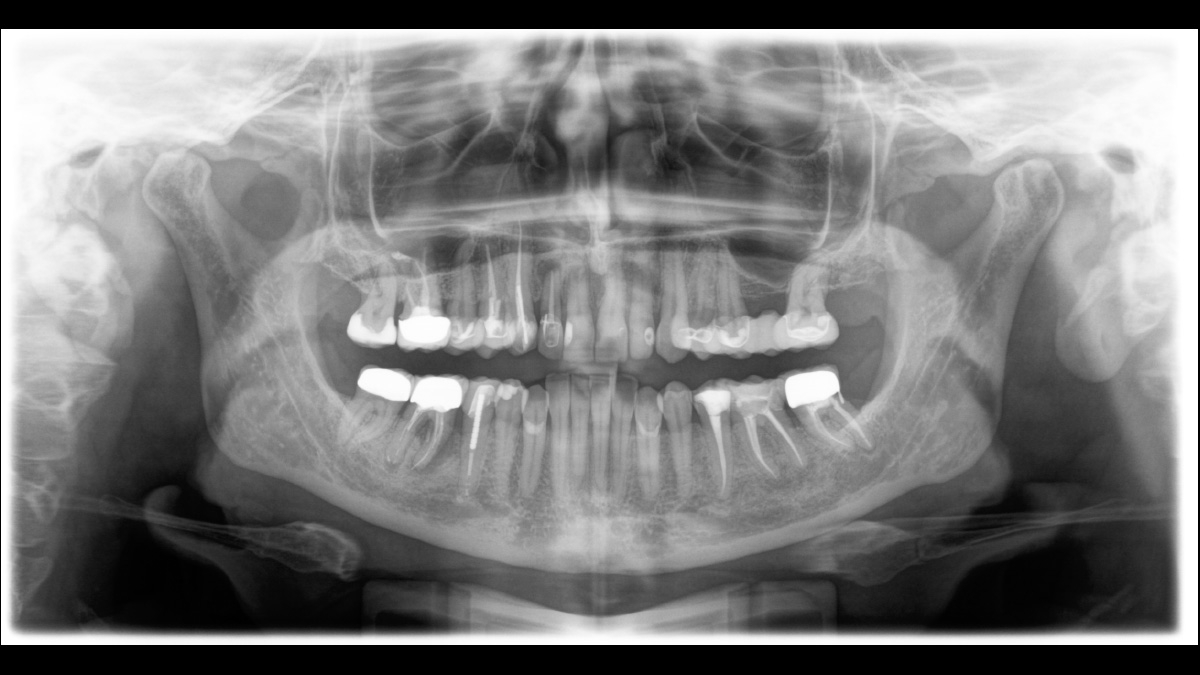
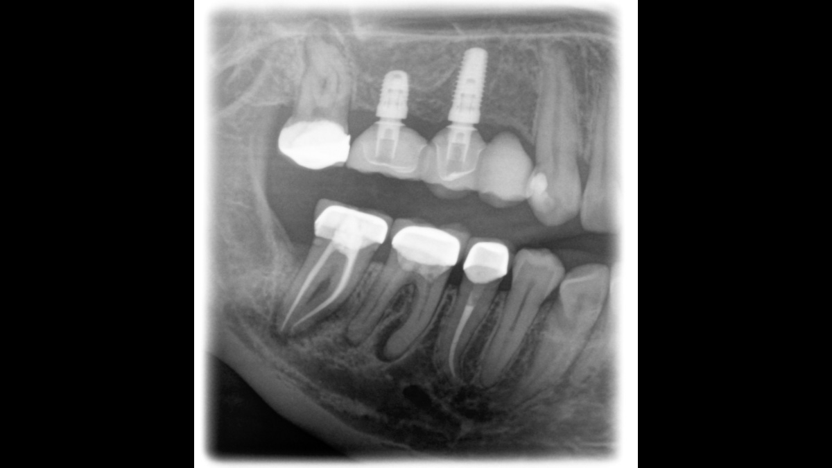
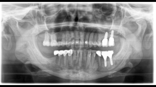
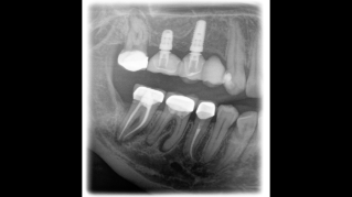
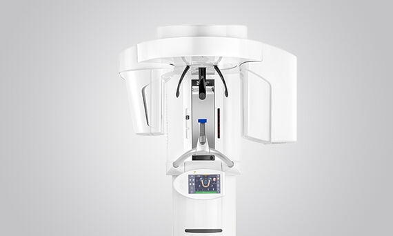
With this high-performance X-ray unit, you have a comprehensive range of capabilities for all of your daily practice tasks. The CsI Plus sensor with autofocus function ensures sharp images, even in anatomically difficult cases.

The 2D All-rounder

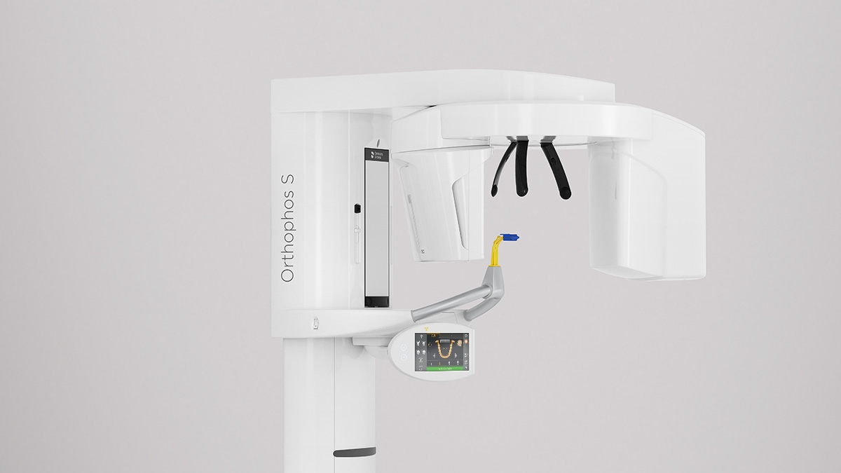
The all-round 2D X-ray device with a comprehensive range of services for every practice. The Orthophos S is a reliable partner and is optimized for everyday tasks: Its CsI Plus sensor with autofocus function ensures clear images, even in anatomically difficult cases. The patented occlusal bite block automatically adjusts the patient’s head tilt to the correct position. For use in orthodontics, the Orthophos S is also available with an optional ceph arm. And because future-proofing is important to Dentsply Sirona,  the system is 3D ready and the cephalometric arm can be retrofitted at any time.


Your Advantages at a Glance

With the Orthophos S 2D, your practice is equipped for the future – with its excellent image quality and 3D upgradability, you’ve got a reliable partner for all of your 2D imaging needs.

Sharp and autofocused images

Even in anatomically difficult cases with the 2D CsI Plus sensor with autofocus function

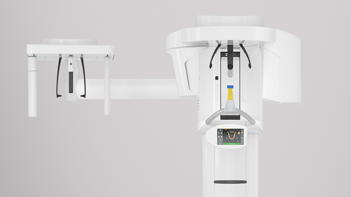
Ceph arm on the left or right

For ceph images, can be ordered as an option or can be retrofitted at any time

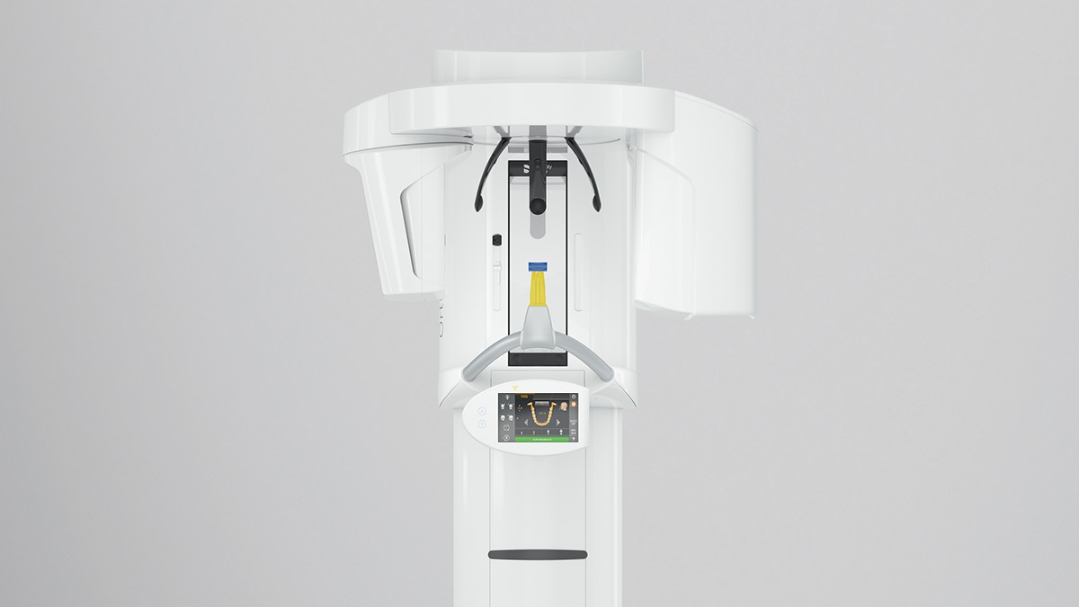
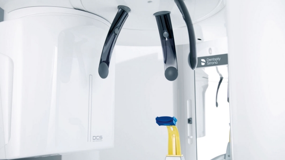
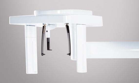
Safe and proven patient positioning

With motorized temple and forehead supports, automatic temple width measurement, light localizers and handles for additional positioning stability

High consistency and reproducibility

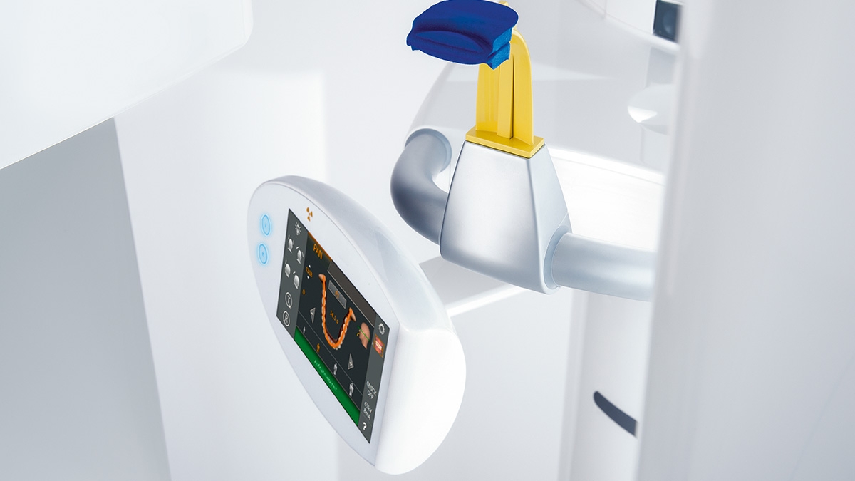
Thanks to automatic patient positioning with the patented occlusal bite block

Orthophos  2D – Highlights


Easy to Use, Safe Positioning

For you, using an x-ray machine is about two things: Getting the best possible image and having your patient feel comfortable. For both, the Orthophos S 2D offers unique, patented solutions: 

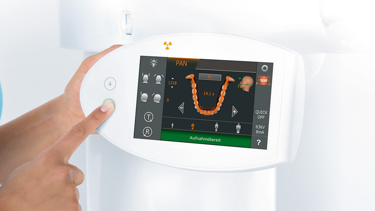
  • The intuitive user interface of the EasyPad
  • Automatic positioning aids such as the patented occlusal bite block and the 3-point head fixation
  • Integrated temple width measurement

Koncepcja pozycjonowania pacjenta

Opracowaliśmy 10-punktową koncepcję ułatwiającą pozycjonowanie pacjenta. Nasza koncepcja to przede wszystkim wysoka jakość obrazu oraz komfort dla pacjenta i asysty. Koncepcja ta wspiera i zapewnia narzędzia potrzebne do wykonania najwyższej jakości obrazów do analizy leczenia. Opatentowana technologia wielofunkcyjnych zagryzaków, na przykład automatycznie ustalający prawidłowe nachylenie głowy pacjenta, pozycjonując go w płaszczyźnie zgryzu wpływa na szybkość i jakość pozycjonowania. Do dyspozycji mamy również 3-punktowe mocowanie głowy wraz z pomiarem anatomii czaszki pacjenta. Wygodne uchwyty dla dłoni zapewniają stabilne pozycjonowanie ograniczając niepotrzebne skany korygujące.

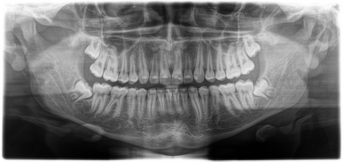
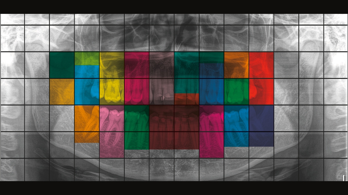
Autofocus – for Excellent Images, automatically

To get a panoramic X-ray image with high definition, the right focus is essential. The jaw must be in the sharp image layer of the device. For this, the Orthophos creates several thousand individual images in one revolution and automatically recognizes the areas in which the jaw is optimally positioned. These are displayed in an overall image with maximum sharpness – without any manual intermediate steps.


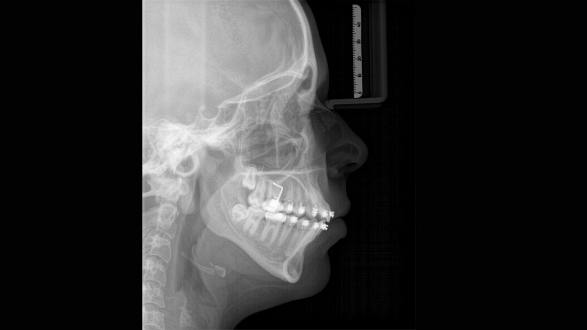
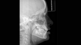
Cephalometric Imaging

Orthophos S 2D offers the capability to install a cephalometric arm at any time. And to be sure it fits in your x-ray room, the arm can be mounted either on the right side or left side of the unit.

Ceph programs that cover all orthodontic needs

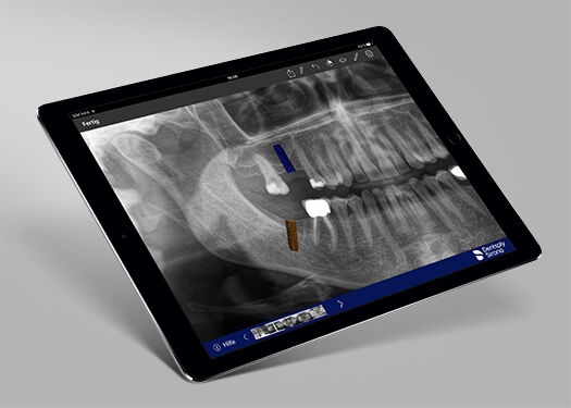
Using a dedicated sensor, you get lateral and symmetrical p.a. or a.p. as well as carpus images. For displaced teeth, you can also use 3D x-rays to determine their exact location.


Technical Data

Performance features
Orthophos SL 2DOrthophos S 2DOrthophos E
X-ray generator
60 - 90 kV, 3-16mA60 - 90 kV, 3-16mA60 - 90 kV, 3-16mA                
Panoramic exposure time
P1: max 14,2 s
P1 Quickshot: max 9,1 s 
P1: max 14,2 s
P1 Quickshot: max 9,1 s
P1: max 14,2 s
Radiation time Ceph
Standard 9,4 s
Quickshot 4,7 s
Standard 9,4 s
Quickshot 4,7 s
Standard 9,4 s
User interface
EasyPadEasyPadMultiPad
Patient positioning
automatic (occlusal bite block) 
manual
Panorama technology
DCSCsI PlusCsI
Autofocus
yesyes-
Ceph arm (optional)
left or rightleft or right
left
Ceph unit with 2 sensors
yes
yes
optional
Quickshot
yes
yes
-
Fields of View
upgradeableupgradeable
-
3D Low Dose
upgradeable
upgradeable
-
HD mode
upgradeable
upgradeable
-
Base
optional
optional
optional
Wheelchair accessible
yesyesyes
Remote control
optional
optional
optional
Ambient Lightyes (backlight)--

All 2D programs at a Glance

ProgrammeOrthophos SL 2DOrthophos S 2DOrthophos E 
Standard panorama image

P1, P2, P10

P1, P2, P10
P1, P10
Image detail left side or right side 

P1, P1A, P1C P2, P2A, P2CP10, P10A, P10C BW1

P1, P1A, P1C P2, P2A, P2CP10, P10A, P10C BW1

P1L, P1R

Image detail individual quadrants 

P1, P1A, P1C P2, P2A, P2CP10, P10A, P10C 

P1, P1A, P1C P2, P2A, P2CP10, P10A, P10C 

-
Image detail upper or lower jaw 

P1, P1A, P1C P2, P2A, P2CP10, P10A, P10C, P12

P1, P1A, P1C P2, P2A, P2CP10, P10A, P10C, P12

-
Constant magnification

P1C, P2C, P10C

P1C, P2C, P10C

P1C

Artifact-reduced 

P1A, P2A, P10A

P1A, P2A, P10A

P1A

Thick layer front 

P12

P12

P12

Sinusoidal images 

S1, S3

S1, S3

S1

Multislice in posterior tooth
--

MS1 

Mandibular joint

TM1.1, TM1.2, TM3

TM1.1, TM1.2, TM3

TM1.1, TM1.2
Bitewing image 

BW1, BW2

BW1, BW2

BW1

Ceph (optional)

C1, C2, C3, C3F, C4

C1, C2, C3, C3F, C4

C1, C2, C3, C3F, C4


FAQ

For more details please see Sidexis 4 system requirements

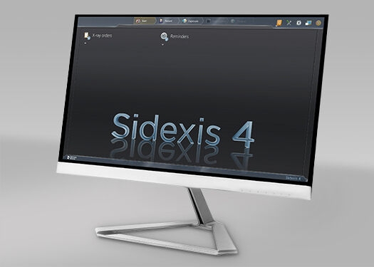
Dentsply Sirona 2D units work exclusively with Sidexis 4. Nevertheless data migration from Sidexis XG to Sidexis 4 is very easy. Sidexis 4 allows for the full digital experience with the latest tools.

The Orthophos SL 2D and the Orthophos S 2D allow a 3D upgrade. Axeos is a dedicated hybrid unit. The Orthophos E does not offer this option.


Rodzina aparatów do obrazowania 2D/3D

Dowiedz się więcej o naszych aparatach 2D oraz hybrydowych aparatach 2D/3D oraz pobierz katalog o rodzinie aparatów zewnątrzustnych Dentsply Sirona.

Dentist with Axeos unit in the background

See More with Certainty

Do more with certainty - Imaging Solutions for Implantology 5 MB

See more with certainty - Imaging Solutions for Orthodontics 2 MB

Find out more about Dentsply Sirona Imaging Solutions for Orthodontics

More Benefits for your Practice

Whether hardware or software, a variety of imaging solutions are waiting for you.


3D Units

Axeos

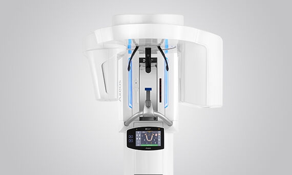
The versatile 2D/3D hybrid unit with a big FOV and high image quality for practices with a wide treatment spectrum. Axeos convinces not only with its high-caliber performance, but also with its focus on comfort and design as well.

Orthophos SL 3D

The high-end 2D/3D hybrid unit features revolutionary technology and patented positioning solutions. An optional ceph arm provides additional benefits, adding to the already broad range of offerings.

Orthophos S 3D

This All-round 2D/3D hybrid unit offers a full range of capabilities to support all of the 2D and 3D tasks in your practice. Thanks to its flexible volume sizes and adjustable dose options, you have a great partner to support your workflows.


Skontaktuj się z nami po więcej informacji

Dowiedz się więcej o systemach obrazowania Dentsply Sirona i poproś o informacje na temat obrazowania wewnątrzustnego, oprogramowania lub technologii obrazowania 2D i 3D.