image-comparison grip-lines arrow-top-right zip arrow-right video minus arrow-back text shop twitter file home quick-order arrow-left youtube search arrow-down check all filter site_references image Slice 1 zoom 🎬 icons / _ icon size / _ download-queue Created with Sketch. chevron-down arrow-right picture pdf podcast exe plus hotline close xxx-1 burger clipboard linked-in ppt Artboard 🎬 icons / _ icon size / _ download-queue_add Created with Sketch. Layer 1 Created by potrace 1.15, written by Peter Selinger 2001-2017 My Account press_releases Created with Sketch. PRESS ds-instruments ds-preventive ds-implants ds-endodontics Cart presentation Created with Sketch. DS-Preventive-Icon-Grey video Created with Sketch. other Created with Sketch. text_piece Created with Sketch. promotion Created with Sketch. Spare Parts ds-imaging-systems case_study Created with Sketch. ds-treatment-centers broschure Created with Sketch. ds-prosthetics ds-restorative video Created with Sketch. flyer Created with Sketch. whitepaper Created with Sketch. article Created with Sketch. image Created with Sketch. Service ds-cad-cam Artboard Quick Order Dentist Chair study Created with Sketch. ds-orthodontics ds-implants ds-endodontics ds-cerec digital-scan grinder ds-diagnosis END-Website-Icons-Shaping-Cleaning-Obturation step_1 analyse ds-abutment-placement more-quality initiative scan more-quality ds-polishing digital ds-irrigation ds-treatment-centers Test treatment ds-restorative ds-implant-planning realistic construct book ds-planning-impression END-Website-Icons-Shaping-Cleaning-Obturation END-Website-Icons-Shaping-Cleaning-Obturation mirror anwender ds-preventive planning-2 ds-obturation ds-cad-cam 04 ds-implant-placement ds-retention-protection ds-anesthetics restauration form ds-inlab oven CORP-icon-grey-screen-tooth-cavity-pulpa robot plan place ds-orthodontics PDF-Formular_ganzneu PDF overlay ds-bone-regeneration ds-treatment-preparation vorher_nachher ds-maging-systems wifi END-Website-Icons-Shaping-Cleaning-Obturation omnicam PDF-Formular_neu_vonGünther treatment consulting planning comparison ds-set-up ds-prosthetics support step_3 ds-tooth-preparation ds-treatments ds-scaling step_2 ds-instruments software quote-english quote-french quote-text-german Created with Sketch. upload Created with Sketch. version Created with Sketch. ds-endodontics globe
Dentsply Sirona Poland
Polski
Home Back

Imaging Systems


Why digital imaging?

With Imaging Systems from Dentsply Sirona you can trust in the safety that more than 120 years of experience and knowledge provide. As the market leader in dental imaging, our solutions provide unparalleled diagnostic image quality at the lowest reasonable radiation dose, while being intuitive and easy to use.

Digital imaging, as opposed to analogue systems, offers many essential benefits for diagnosis and patient workflow in a clinic. Additionally, it facilitates dental education, since students are able to work with the comprehensive digital record of the patient.

Benefits for education

Comprehensive treatment planning

All digital images and patient data are stored in the digital record improving the quality of the diagnosis and facilitating treatment planning – even when the patient isn’t present.

Learning with state-of-the art devices

With Dentsply Sirona imaging solutions students learn with the latest technology from the start. They become familiar with correct patient positioning and exposure techniques, as well as with an efficient treatment and patient flow.

Benefits for diagnosis and treatment flow

Better image quality

Digital X-ray imaging offers an image resolution that guarantees large image formats and high quality. The result: high-resolution images with optimum diagnostic reliability, which leaves no question unanswered and makes all relevant structures visible.

Faster results

Radiography with film processing is time consuming and requires many steps. Digital images can be displayed immediately, which gives you more time with the patient and shortens treatment appointments.

Improved patient comfort

Digital imaging is more pleasant for patients because they can receive their diagnosis directly after the image has been taken. Digital X-ray imaging also reduces waiting times.

More flexibility

Digital images do not have to be printed and stored, they are automatically transferred into the digital record of the patient. The image can then be discussed with the patient in the treatment room or accessed remotely, allowing for consultation with other colleagues outside of the practice.


More than imaging – real solutions

To ensure maximum insight and confidence 

As a pioneer in dental Imaging Systems, we continue to set the standard in modern, digital imaging. Our solutions provide unparalleled diagnostic image quality at the lowest reasonable radiation dose, while being intuitive and easy to use. They can be used to support workflows in all dental disciplines, integrating imaging systems and software to enable truly better, safer, and faster procedures.

Our intraoral range consists of X-ray tubes, a family of sensors, a phosphor plate scanner, and a complete range of accessories for holding and positioning sensors and phosphor plates.

Our range of extra oral imaging systems begins with a proven entry level 2D unit, extends to a hybrid 2D/3D unit, and goes all the way to systems with 3D, and large field of view CBCT for full face scanning.

Imaging software includes solutions for both generalists and specialists, enabling them to provide better and more integrated care in disciplines like implantology, endodontics, function diagnostics and sleep apnea.


3D imaging

Experience the difference

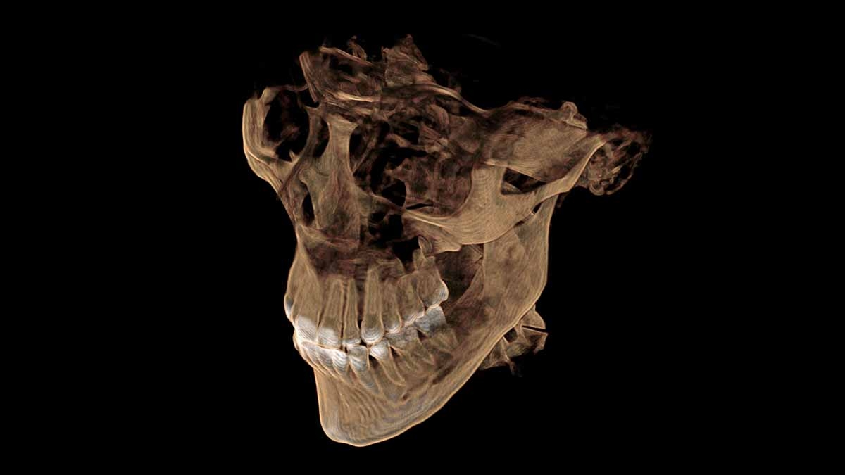
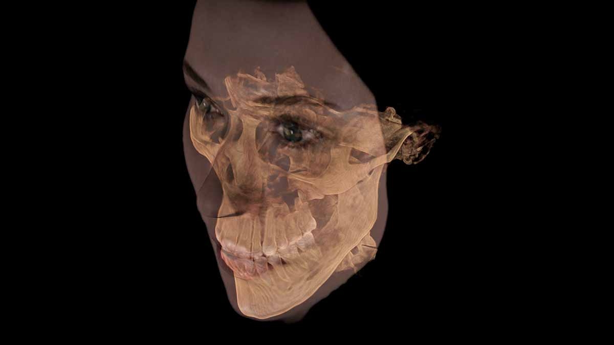
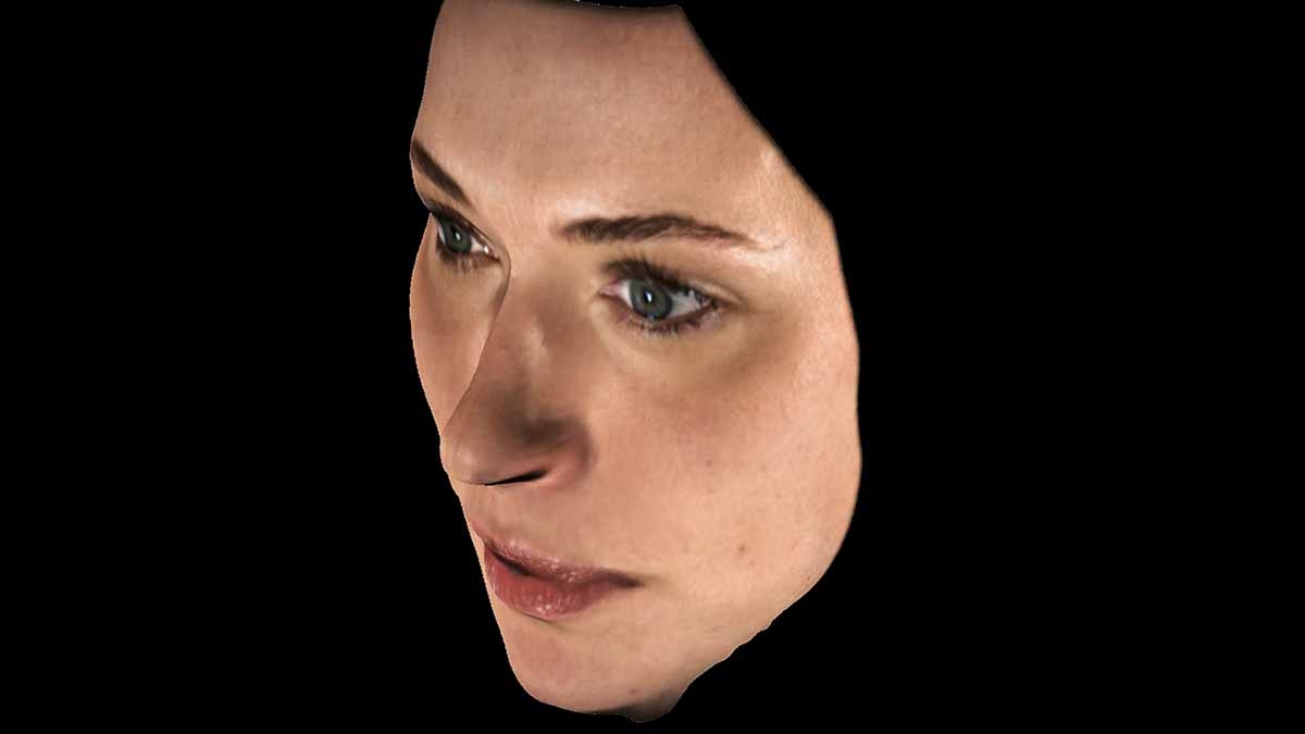
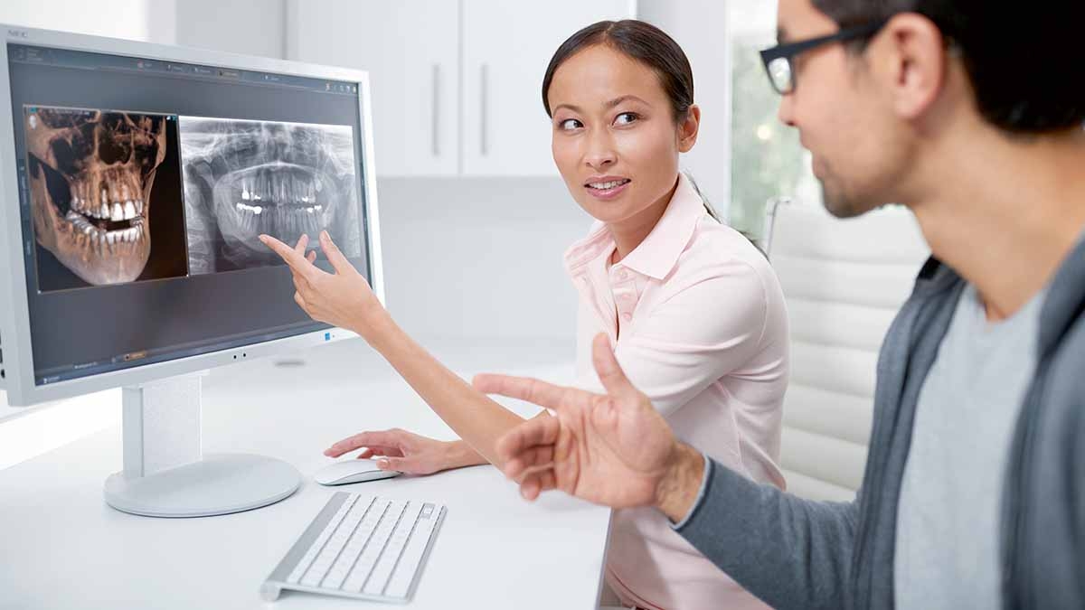
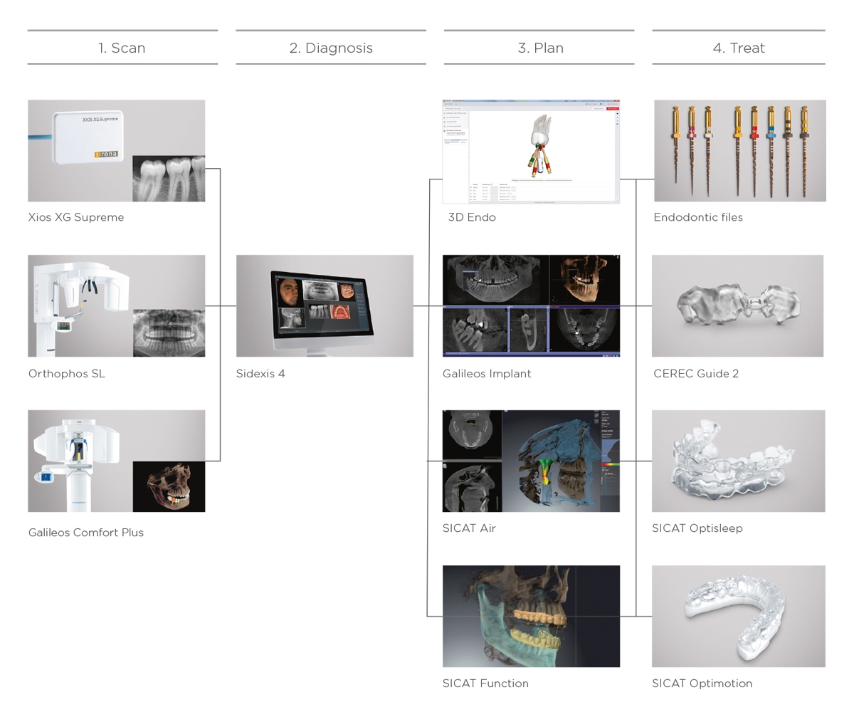
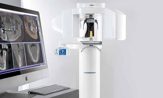
3D imaging has revolutionized dentistry. With its capability to see any given situation from new and different perspectives, its technology has introduced a new layer of safety and efficiency to clinics and practices worldwide. 3D imaging enables completely new dimensions in diagnostics. Today, certain dental specializations would not be possible without 3D imaging. Guide your students through this fascinating world with Dentsply Sirona’s dedicated CBCT unit Galileos Comfort Plus.

3D Low Dose Mode

The safety of 3D images at the dose level of 2D X-rays. This is what the Low Dose Mode offers for a large number of indications.

What advantages does 3D X-ray technology offer?

Expanded diagnostic capability

Thanks to the precise imaging technique, you have additional options for diagnosing: the path of jaw and facial nerves, the positional relationship the teeth, the state of the bone, and tooth and jaw diseases.

Greater diagnostic accuracy

3D X-ray images ensure precise diagnostics, even for special and complex situations. For example, the 3D X-ray image shows a tooth root from all angles and exposes hidden changes.

Communication with the patient

Diagnoses and treatment planning can be communicated far more easily to patients with 3D images. This enables them to better understand treatment recommendations and to accept them more readily.

Larger range of services

With 3D technology, you offer your patients a larger range of services and thus enhance the sustainability of your clinic.


Facescan

The Facescan plots the patient’s facial surfaces at the same time as the X-ray image is being taken. With a realistic image of their own face, patients understand and accept treatment recommendations more readily.