image-comparison grip-lines arrow-top-right zip arrow-right video minus arrow-back text shop twitter file home quick-order arrow-left youtube search arrow-down check all filter site_references image Slice 1 zoom 🎬 icons / _ icon size / _ download-queue Created with Sketch. chevron-down arrow-right picture pdf podcast exe plus hotline close xxx-1 burger clipboard linked-in ppt Artboard 🎬 icons / _ icon size / _ download-queue_add Created with Sketch. Layer 1 Created by potrace 1.15, written by Peter Selinger 2001-2017 My Account press_releases Created with Sketch. PRESS ds-instruments ds-preventive ds-implants ds-endodontics Cart presentation Created with Sketch. DS-Preventive-Icon-Grey video Created with Sketch. other Created with Sketch. text_piece Created with Sketch. promotion Created with Sketch. Spare Parts ds-imaging-systems case_study Created with Sketch. ds-treatment-centers broschure Created with Sketch. ds-prosthetics ds-restorative video Created with Sketch. flyer Created with Sketch. whitepaper Created with Sketch. article Created with Sketch. image Created with Sketch. Service ds-cad-cam Artboard Quick Order Dentist Chair study Created with Sketch. ds-orthodontics ds-implants ds-endodontics ds-cerec digital-scan grinder ds-diagnosis END-Website-Icons-Shaping-Cleaning-Obturation step_1 analyse ds-abutment-placement more-quality initiative scan more-quality ds-polishing digital ds-irrigation ds-treatment-centers Test treatment ds-restorative ds-implant-planning realistic construct book ds-planning-impression END-Website-Icons-Shaping-Cleaning-Obturation END-Website-Icons-Shaping-Cleaning-Obturation mirror anwender ds-preventive planning-2 ds-obturation ds-cad-cam 04 ds-implant-placement ds-retention-protection ds-anesthetics restauration form ds-inlab oven CORP-icon-grey-screen-tooth-cavity-pulpa robot plan place ds-orthodontics PDF-Formular_ganzneu PDF overlay ds-bone-regeneration ds-treatment-preparation vorher_nachher ds-maging-systems wifi END-Website-Icons-Shaping-Cleaning-Obturation omnicam PDF-Formular_neu_vonGünther treatment consulting planning comparison ds-set-up ds-prosthetics support step_3 ds-tooth-preparation ds-treatments ds-scaling step_2 ds-instruments software quote-english quote-french quote-text-german Created with Sketch. upload Created with Sketch. version Created with Sketch. ds-endodontics globe
Dentsply Sirona China
中文
主页 后退

2D 影像

Orthophos 2D 影像系列能够为诊所提供相应的解决方案。

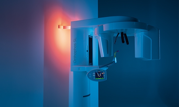
Orthophos SL、Orthophos XG 的 2D 设备

Orthophos 2D 影像系列能够为诊所提供相应的解决方案。从进入数字放射影像到专业领域的出色解决方案 - 登士柏西诺德拥有精心设计的产品系列,可满足您的需求。Orthophos 2D 影像设备随登士柏西诺德影像软件 Sidexis 4 一起提供,交付更好和更快速的诊断。


登士柏西诺德 2D 影像组合一览

您可以选择两种不同的型号,从入门级的数字影像设备一直到卓越的集成 2D 影像设备。

Orthophos XG 3

设备操作简单,可无缝集成到您的诊所日常工作之中,为您节省宝贵的时间。

Orthophos XG 5

口腔正畸医生的专业合作伙伴,拥有专门的儿科和头颅侧位规划,其中头颅侧位臂是可选的。

优势

优质的影像质量,实现高效的诊断

始终遵循 ALARA 原则,我们的 2D 影像系列确保您可以在尽可能低的剂量下针对给定的适应症,得到所需的影像质量。

凭借经过验证的质量,让您的诊所可靠运行

凭借骄人的“德国制造”,全球销售量超过 100,000,我们可以肯定地说:Orthophos 设备久经验证。

易于操作,重现性好

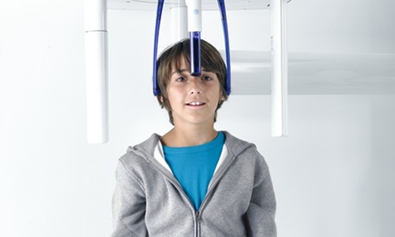
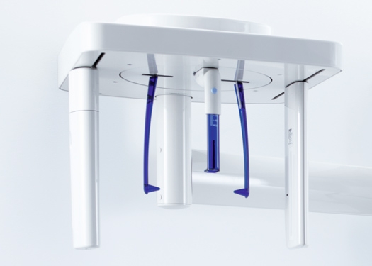
稳定的患者定位系统以及直观的控制面板旨在将学习曲线降至最低,并支持您的诊所效率。

数字优势,让您的生活更轻松

无需暗室,无需化学品,运行成本更低,剂量更少,工作流程更快。这些只是我们的数字 2D 系列为您的诊所带来的一部分益处。


2D 影像系列的特性亮点

患者定位:充分利用可重现影像质量的支柱

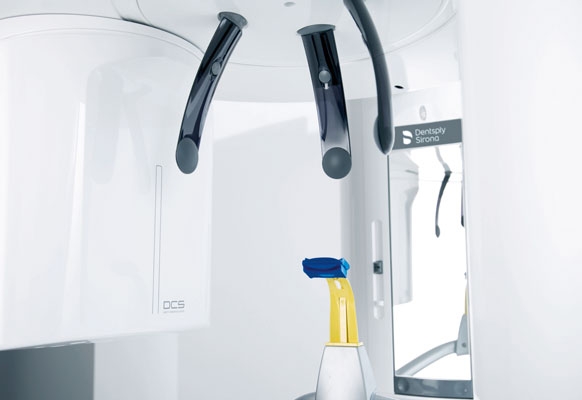
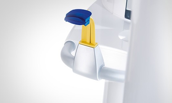
登士柏西诺德咬合块安全操作,易于定位,高影像质量,可自动确定患者头部的正确倾斜度,使患者在咬合面快速定位,配合 3 点头部固定和牢固的手柄,以确保稳定定位,避免不必要的校正扫描。


2D 全景影像

尽管 3D 影像在越来越多的牙科诊所中日渐成熟,但在许多情况下,2D 影像仍然具有应有的地位,这主要是由于放射卫生学的原因。它是现代牙科诊断的核心组成部分。在影像质量的改进方面,2D 技术中可用的选项远远没有用尽。来自登士柏西诺德的 2D 影像设备在日常处理方面得到了积极的评价,这要归功于高质量的清晰影像和操作的技术创新。
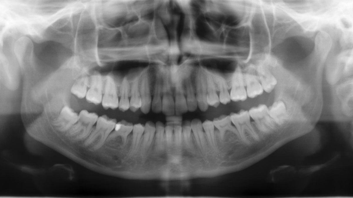
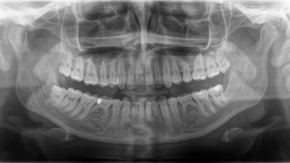
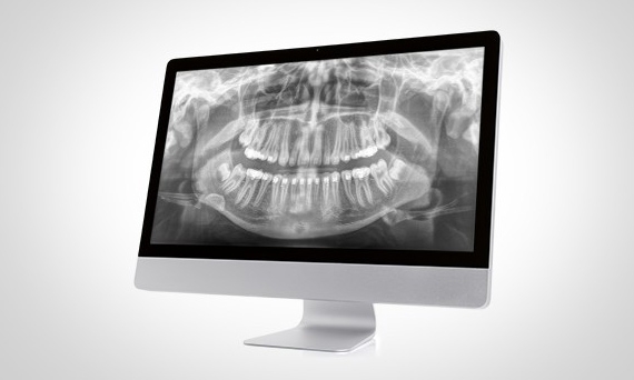
全景影像质量不断发展

左侧:10 年的 PAN 影像。右侧:用 Orthophos SL 拍摄的 DCS 影像


头颅侧位影像

Orthophos SL 2D、Orthophos XG 3D ready 和 Orthophos XG 5 可安装头颅侧位臂。此外,为了确保它适合您的影像室,头颅侧位臂可以安装在设备的右侧或左侧(Orthophos XG 5 仅限左侧)。

头颅侧位程序涵盖所有口腔正畸学需求

使用专用传感器,您可以获得横向和对称的 p.a. 或者 a.p.,以及 carpus 影像。

相关软件

ASTRA

ASTRA 软件算法提高了 2D 全景和头颅侧位影像的质量。

自动定位

咬合块简化了患者的定位。

ALARA

使用尽可能低剂量获得更佳影像质量 - 遵循 ALARA 原则。

技术数据

概览表

2D 设备Orthophos XG (2D)Orthophos SL 2D
型号  
 Orthophos XG 3:7 个程序,无头颅侧位选项,无 3D 升级
Orthophos SL 2D:XX 程序,右侧或左侧头颅侧位臂选项,可 3D 升级
Orthophos XG 5:XX 程序,右侧头颅侧位臂选项,无 3D 升级
Orthophos XG 3Dready:XX 程序,右侧或左侧头颅侧位臂选项,可 3D 升级
球管
 
X 射线发生器
60-90 kV; 3-16 mA
焦点尺寸符合 IEC 60336
0.5 mm
总过滤率符合 IEC 60522
> 2.5 mm Al
2D 影像
 
传感器

数字 CCD 线传感器,

可重新插入

数字碲化镉 (CdTe) 传感器

采用直接转换传感器 (DCS) 技术

有源传感器区域
138 x 6.48 mm146 x 6 mm
像素大小
27 μm
100 μm
聚焦传感器距离
530 mm
524 mm

头颅侧位影像

(可选)


左侧或右侧
传感器
采用 CCD 技术的数字线传感器(如果 XG 型号可重新插入)
有源传感器区域
230 x 6.48 mm
像素大小
27 μm
聚焦传感器距离
1,714 mm
一般信息
 
原产地
德国制造。由登士柏西诺德制造。
尺寸(长 × 宽 × 高)
无头颅侧位:1.411 × 1.28 × 2.25 m / 5'3'' × 4'2.4'' × 7' 4.5''
带头颅侧位:1.411 × 2.155 × 2.25 m / 5'3'' × 7'0.6'' × 7' 4.5'' 
重量无头颅侧位:110 kg / 242 lbs
带头颅侧位:132 kg / 291 lbs
运行条件
 
空气湿度10% 至 95%
温度+10° to +40°C (50° 至 104°F)+18° to +31°C (64° 至 88°F)

常见问题解答

这些要求实际上遵循影像处理软件 Sidexis 4 的要求。关于详细信息,请参阅 Sidexis 4 系统要求

Orthophos SL 仅使用 Sidexis 4。然而,从 Sidexis XG 到 Sidexis 4 的数据迁移非常简单。使用 Orthophos XG 设备,您可以根据需要继续使用 Sidexis XG,尽管我们建议使用最新工具换到 Sidexis 4,以获得完整的数字体验。


联系我们

了解有关登士柏西诺德影像系统的更多信息,并索取有关口内图像、软件或 2D 和 3D 影像技术的产品信息。