
Give us a call or provide your contact details below, and a Dentsply Sirona representative will be in touch soon.

Contact Us
Take your practice to the next level.
Discover more ways to bring innovation to your practice.

Position Yourself for Success
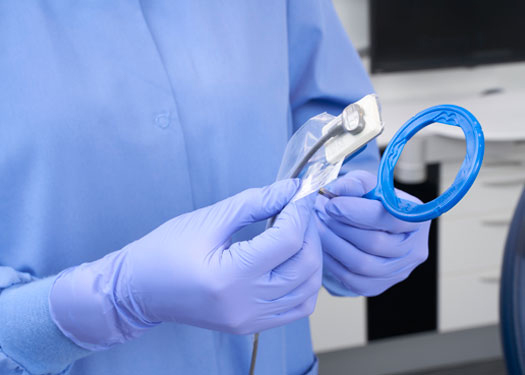
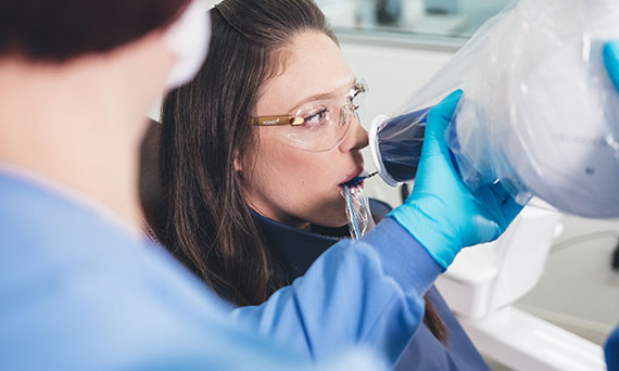
Capturing an excellent intraoral X-ray starts with proper sensor positioning.
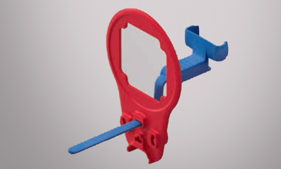
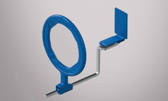
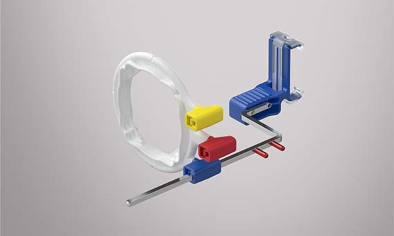
Our Dentsply Sirona Xios Intraoral Holder Systems help ensure a comfortable experience for your patient and provide an exceptional tool to support your dental assistant or hygienist in capturing a precise intraoral X-ray safely and efficiently. We offer multiple options to select from with our Xios AimRight Sensor Holder Systems and Xios Sensor Holder System.
Your Xios Intraoral Holder Systems may quickly become your favorite tools to work with. Designed for intraoral imaging precision and consistency, you’re positioned for success.
Xios Intraoral Holder Systems from Dentsply Sirona
Benefits
Why choose Xios Intraoral Holder Systems?


Image Accuracy
Xios Intraoral Holder Systems help to simplify your diagnostic workflow and provide additional positioning control for an accurate and precise image capture.
Easy Positioning
Holders help to accurately capture the target tooth of interest and add additional stability, streamlining the workflow process.
Safety
Capture diagnostically acceptable intraoral images in a single, well-executed take, maximizing patient comfort and safety.
Positive Patient Experience
Consistent X-rays with minimal added bulk to the holders, and a great tool to help limit image retakes and cut down additional treatment time.

The Intraoral Imaging Workflow
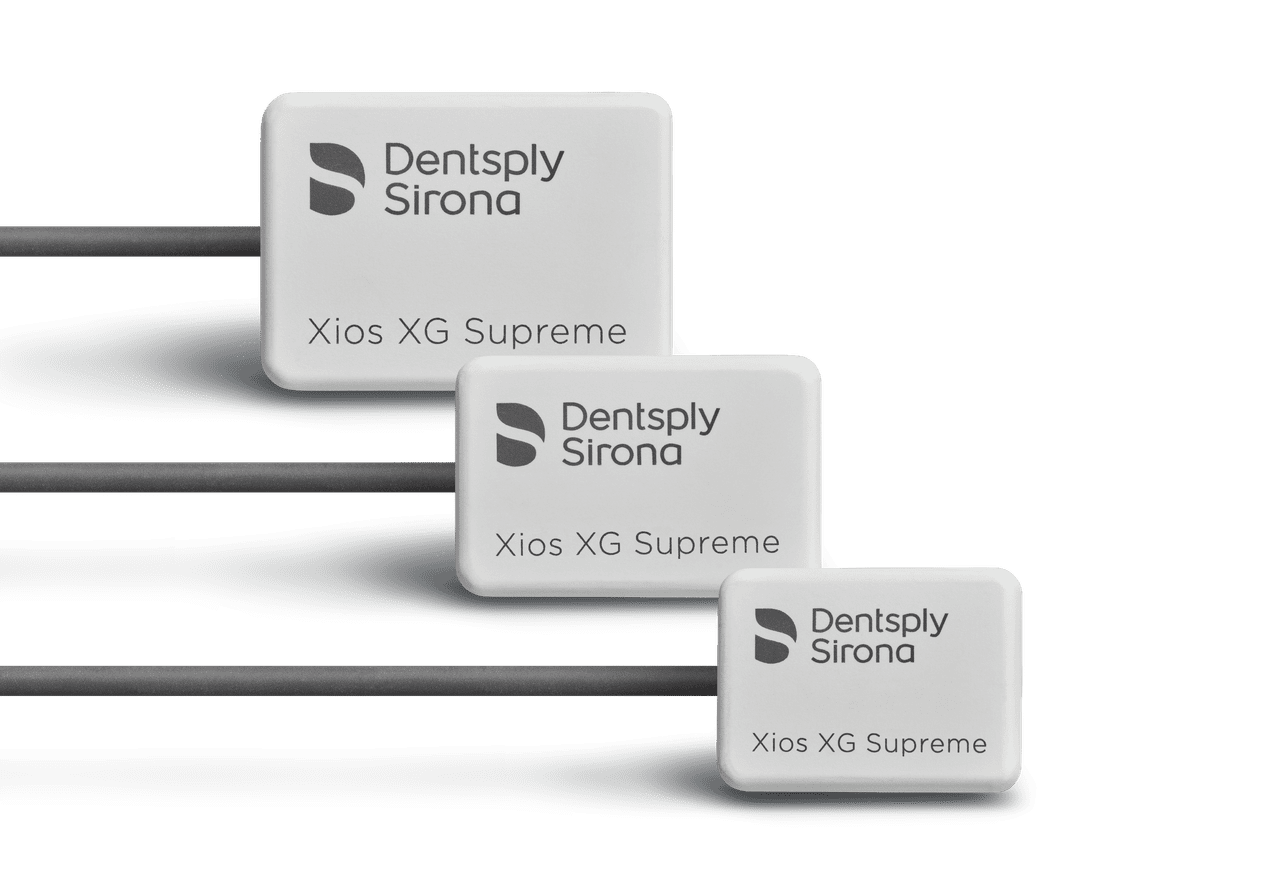
The Xios intraoral imaging accessories from Dentsply Sirona provide industry-leading solutions for all of your intraoral radiography needs. They simplify the diagnostic workflow, helping clinicians work more efficiently while keeping patients comfortable and safe. They provide accurate positioning and control to consistently deliver diagnostically acceptable images in either digital or traditional media. Dentsply Sirona also offers additional options with our Rinn holder system product portfolio. No matter which way you want to work, we have the tools to support you.
"The ease of positioning means I expose better quality images for my doctors, meaning my patients are not re-exposed to additional radiation. The quality, comfort and safety of the Dentsply Sirona sensor make it an essential tool in the provision of elevated and comprehensive patient care."
Katrina Sanders, RDH, Phoenix, AZ, USA
The ALARA Principal — As low as reasonably achievable
Image quality and radiation exposure matter.
The need for clear, diagnostically acceptable images should never take priority over every clinician’s first principle: do no harm. That means keeping radiation exposure for your staff and patients as low as reasonably achievable: the ALARA principle.
Every inaccurate image requires a retake and the more images retaken, the more the clinician and the patient are exposed to ionizing radiation. Your goal is to capture diagnostically acceptable intraoral images in a single, well-executed take, maximizing patient comfort and safety while minimizing chair time.
The most common reasons for intraoral radiography retakes are due to improper positioning, angulation, and media placement.1
Tips and education for better intraoral imaging
The goal of dental radiology is to obtain high-quality images while minimizing radiation exposure and keeping every patient as comfortable as possible. Consider how these tips may assist in challenging situations.
PARALLELING VS BISECTING
- Know what to choose based on anatomical challenge.
- The paralleling technique using aiming devices is recommended for fewer errors and retakes.
- The bisecting angle technique using media holders are recommended for patients with anatomical challenges, and during endodontic and implant procedures.
SENSOR SELECTION
- Use the largest sensor possible to obtain maximum information.
- Have a protocol in place for when to switch to a smaller sensor.
GENERAL SENSOR POSITIONING
- Place in the center of the mouth for the paralleling technique.
- Use cotton rolls, especially in vertical positions and edentulous areas.
SENSOR POSITIONING FOR BITEWINGS
- Place the sensor parallel to the teeth.
- Use the indicator slot to align with interproximal spaces on bitewings.
- Align the central beam through the contact areas.
- Adjust vertical angulation on bitewings to capture adequate bone levels.
- With premolar bitewings, to capture the distal of the canine, adjust the media approximately 15 degress distomesial.
1. Acharya S, Pai K, Acharya S. Repeat film analysis and its implications for quality assurance in dental radiology: An institutional case study. Contemporary Clinical Dentistry. 2015;6(3):392-395.
