Orthophos S

image-comparison grip-lines arrow-top-right zip arrow-right video minus arrow-back text shop twitter file home quick-order arrow-left youtube search arrow-down check all filter site_references image Slice 1 zoom 🎬 icons / _ icon size / _ download-queue Created with Sketch. chevron-down arrow-right picture pdf podcast exe plus hotline close xxx-1 burger clipboard linked-in ppt Artboard 🎬 icons / _ icon size / _ download-queue_add Created with Sketch. Layer 1 Created by potrace 1.15, written by Peter Selinger 2001-2017 My Account press_releases Created with Sketch. PRESS ds-instruments ds-preventive ds-implants ds-endodontics Cart presentation Created with Sketch. DS-Preventive-Icon-Grey video Created with Sketch. other Created with Sketch. text_piece Created with Sketch. promotion Created with Sketch. Spare Parts ds-imaging-systems case_study Created with Sketch. ds-treatment-centers broschure Created with Sketch. ds-prosthetics ds-restorative video Created with Sketch. flyer Created with Sketch. whitepaper Created with Sketch. article Created with Sketch. image Created with Sketch. Service ds-cad-cam Artboard Quick Order Dentist Chair study Created with Sketch. ds-orthodontics ds-implants ds-endodontics ds-cerec digital-scan grinder ds-diagnosis END-Website-Icons-Shaping-Cleaning-Obturation step_1 analyse ds-abutment-placement more-quality initiative scan more-quality ds-polishing digital ds-irrigation ds-treatment-centers Test END-Website-Icons-Shaping-Cleaning-Obturation treatment ds-restorative ds-implant-planning realistic construct book ds-planning-impression END-Website-Icons-Shaping-Cleaning-Obturation END-Website-Icons-Shaping-Cleaning-Obturation mirror anwender ds-preventive planning-2 ds-obturation ds-cad-cam 04 ds-implant-placement ds-retention-protection ds-anesthetics restauration form ds-inlab oven CORP-icon-grey-screen-tooth-cavity-pulpa robot plan place ds-orthodontics PDF-Formular_ganzneu PDF overlay ds-bone-regeneration ds-treatment-preparation vorher_nachher ds-maging-systems wifi END-Website-Icons-Shaping-Cleaning-Obturation omnicam PDF-Formular_neu_vonGünther treatment consulting planning comparison ds-set-up ds-prosthetics support step_3 ds-tooth-preparation ds-treatments ds-scaling step_2 ds-instruments software quote-english quote-french quote-text-german Created with Sketch. upload Created with Sketch. version Created with Sketch. globe EndoEquipmentGreypng Imaging Grey 2svg EndoGlideShapingGreypng irrigation blue Imaging 1svg Imaging 2svg DS-Preventive-Icon-Grey Final_Implants_Category_Implant-Systems Access Blue obturation blue restoration restoration grey Access Grey treatment EndoEquipmentBluepng irrigation grey Diagnosis primeprint-grey Hub Bluesvg Final_Implants_Procedure_Single-tooth Diagnsis blue Imaging blue EndoRetretmentBluepng restoration blue Final_Implants_Procedure_Edentulous-therapy Final_Implants_Procedure_Hard-and-soft-tissue-management obtruation grey endo grey treatment blue Imaging blue 1svg Imaging grey EndoGlideShapingBluepng EndoRetretmentGreypng polishing Hubsvg Final_Implants_Category_Implant-Dentistry polishing blue Diagnosis Grey Cerec Bluesvg Imaging Blue 2svg treatment grey Hub Greysvg Scaling Cerec Greysvg endo blue polishing grey search Imaging Grey 1svg scaling blue Cerecsvg DS Points Logo Imaging scaling grey

A true all-rounder of its class

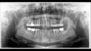
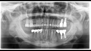
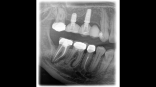
The high-quality 2D/3D X-ray device with a comprehensive range of services for every practice. Whether as a pure 2D device or including a 3D module – the Orthophos S is a reliable partner and optimized for everyday tasks. Its CsI Plus sensor with autofocus function ensures clear images, even in anatomically difficult cases. The automatic patient positioning together with the patented occlusal bite block enables an easy and timesaving patient positioning. For use in orthodontics, the Orthophos S is also available with an optional ceph arm. And because future-proofing is important to Dentsply Sirona, the cephalometric arm can be retrofitted at any time.


Why choose the Orthophos S?

Sharp and autofocused images

Even in anatomically difficult cases with the 2D CsI sensor with autofocus function

Selectable volume sizes

From Ø 5 cm x 5.5 cm to 8 cm x 8 cm or optional up to 11 cm x 10 cm

An optional cephalometric arm

The ceph arm can be added on the right or left side of the unit and ordered at the time of purchase or retrofitted at any time

Maximum consistency and Easy reproducibility

Thanks to automatic patient positioning with the patented occlusal bite block

3D according to your needs

With the 3D Low Dose mode, you get 3D images in the dose range of a 2D X-ray. In HD mode (up to 1,400) individual images are captured during a single rotation and converted into a 3D volume with up to 80 μm for low-noise images in high resolution.

Safe and proven patient positioning

With motorized temple and forehead supports, automatic temple width measurement, light localizers and integrated handles for additional positioning stability


Easy to Use. Precise Patient Positioning.

For most practices, they use an X-ray device with two main goals in mind: Capture the best possible image to support an accurate and precise diagnosis and ensure that the patient is comfortable during the process. The Orthophos S offers unique, patented solutions to help support both of these goals with:

  • The EasyPad and its intuitive user interface
  • Automatic positioning aids such as the patented occlusal bite block and the 3-point head fixation
  • Integrated temple width measurement

Autofocus - Exceptional images, no manual steps

The right focus is crucial for excellent panoramic radiographs. With the autofocus function you will automatically receive an image with the best possible sharpness in focus. The Orthophos S creates several thousand individual images in one revolution and automatically recognizes the areas in which the jaw is optimally positioned. A sharp panoramic image is created from the individual projections – without any manual intermediate steps.


Volumes for a variety of indications

A broad range of volume sizes to support your various diagnostic and clinical needs from Ø 5 x 5.5 cm to Ø 11 x 10 cm


Frequently Asked Questions

 The requirements follow those for image processing software Sidexis 4 and Axeos. For more details please see  Sidexis 4 system requirements and Orthophos S installation requirements.

Dentsply Sirona x-ray units work exclusively with Sidexis 4. Nevertheless data migration from Sidexis XG to Sidexis 4 is very easy. Sidexis 4 allows for the full digital experience with the latest tools.

Orthophos S 2D allows a 3D upgrade. Axeos is a dedicated hybrid unit. The Orthophos E does not offer this option.


Contact us for more information

Find out more about the imaging systems from Dentsply Sirona and request information on intraoral imaging, software or 2D and 3D imaging technology.