Contact Us
Take your practice to the next level.
Discover more ways to bring innovation to your practice.
A true all-rounder of its class
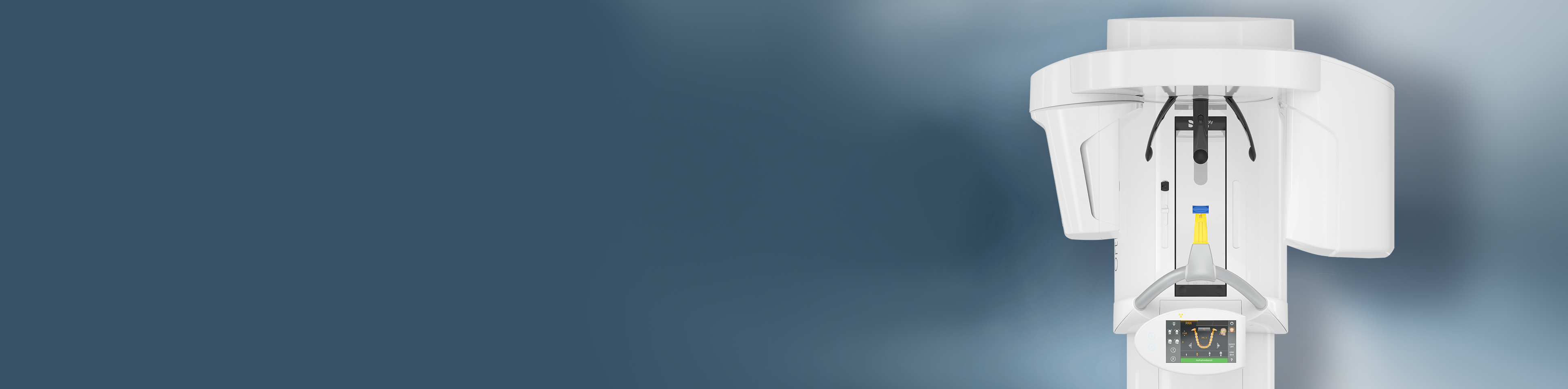
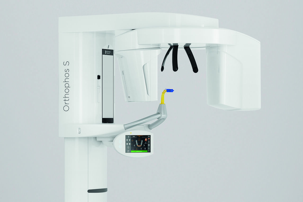
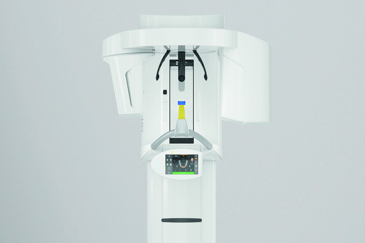
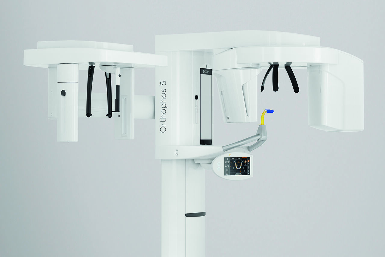
The high-quality 2D/3D X-ray device with a comprehensive range of services for every practice. Whether as a pure 2D device or including a 3D module – the Orthophos S is a reliable partner and optimized for everyday tasks. Its CsI Plus sensor with autofocus function ensures clear images, even in anatomically difficult cases. The automatic patient positioning together with the patented occlusal bite block enables an easy and timesaving patient positioning. For use in orthodontics, the Orthophos S is also available with an optional ceph arm. And because future-proofing is important to Dentsply Sirona, the cephalometric arm can be retrofitted at any time.
Orthophos S from Dentsply Sirona

Benefits
Why choose the Orthophos S?


Sharp and autofocused images
Even in anatomically difficult cases with the 2D CsI sensor with autofocus function
Selectable volume sizes
From Ø 5 cm x 5.5 cm to 8 cm x 8 cm or optional up to 11 cm x 10 cm
An optional cephalometric arm
The ceph arm can be added on the right or left side of the unit and ordered at the time of purchase or retrofitted at any time
Maximum consistency and Easy reproducibility
Thanks to automatic patient positioning with the patented occlusal bite block
3D according to your needs
With the 3D Intelligent Low Dose mode, you get 3D images in the dose range of a 2D X-ray. In HD mode (up to 1,400) individual images are captured during a single rotation and converted into a 3D volume with up to 80 μm for low-noise images in high resolution.
Safe and proven patient positioning
With motorized temple and forehead supports, automatic temple width measurement, light localizers and integrated handles for additional positioning stability
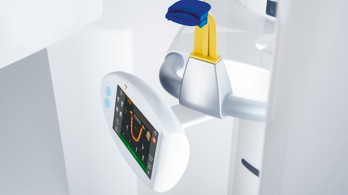
Easy to Use. Precise Patient Positioning.
For most practices, they use an X-ray device with two main goals in mind: Capture the best possible image to support an accurate and precise diagnosis and ensure that the patient is comfortable during the process. The Orthophos S offers unique, patented solutions to help support both of these goals with:
- The EasyPad and its intuitive user interface
- Automatic positioning aids such as the patented occlusal bite block and the 3-point head fixation
- Integrated temple width measurement
Autofocus
Excellent images, without any manual steps
The right focus is crucial for excellent panoramic radiographs. With the autofocus function you will automatically receive an image with the best possible sharpness in focus. Dentsply Sirona Imaging devices take several thousand individual images in one cycle and automatically identify the areas where the jaw is optimally positioned. Without any additional manual steps, these images are then displayed in a final sharp image.

Without Autofocus
Only some parts of the image are in focus, while other areas are blurred.

With Autofocus
The system detects the relevant areas from several thousand individual images in one cycle and automatically identifies the areas where the jaw is optimally positioned.

The result
Sharp images.
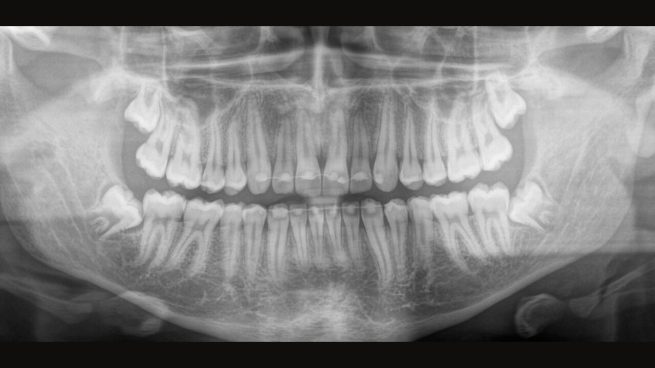
Gallery of 2D Images
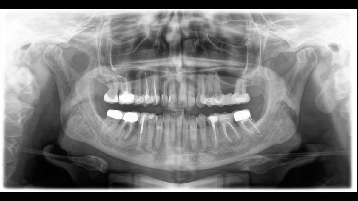
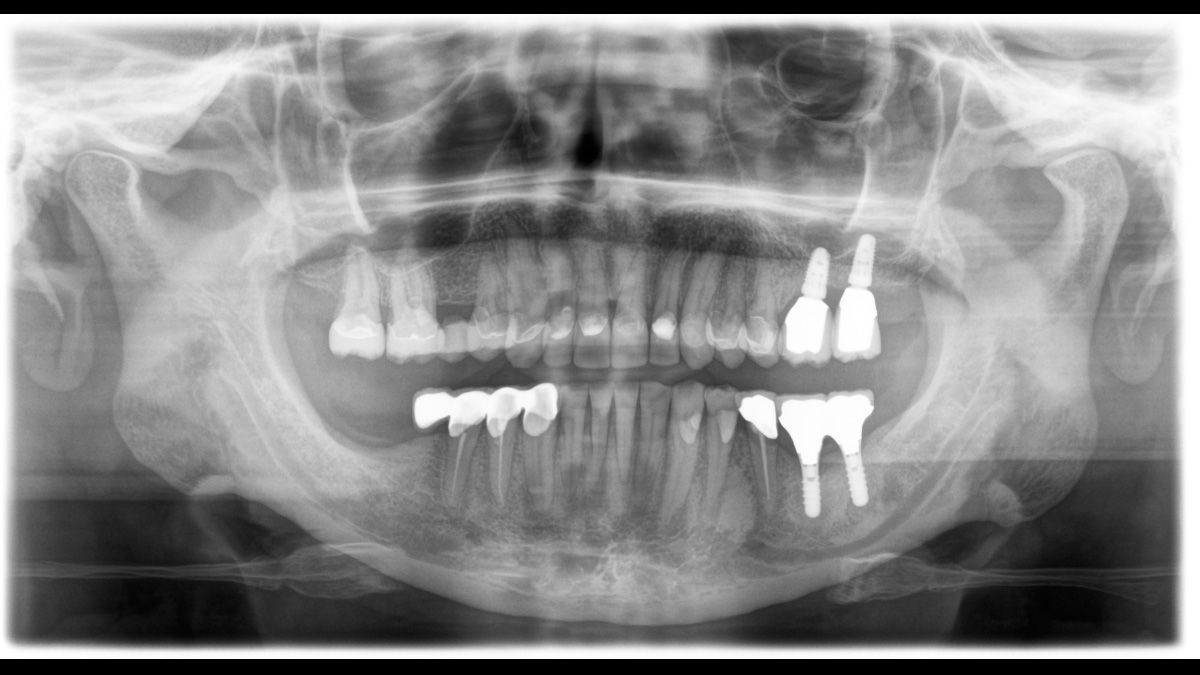
Volumes for a variety of indications
A broad range of volume sizes to support your various diagnostic and clinical needs from Ø 5 x 5.5 cm to Ø 11 x 10 cm
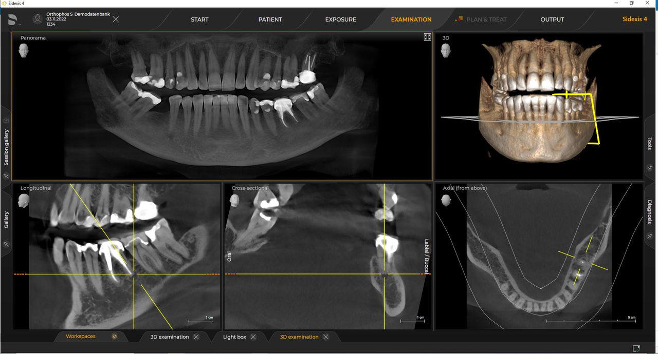
Gallery of 3D Images
 Ø 11 cm x 10 cm HD
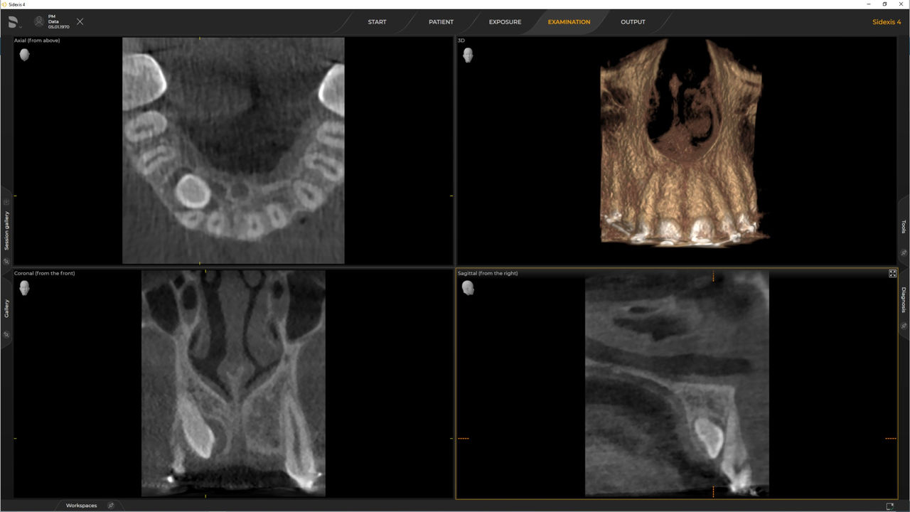 Ø 5 cm x 5.5 cm Low Dose
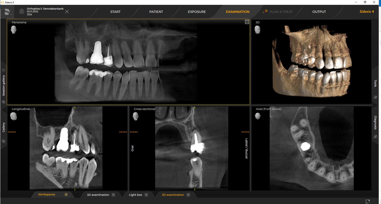 Ø 5 cm x 5.5 cm HD
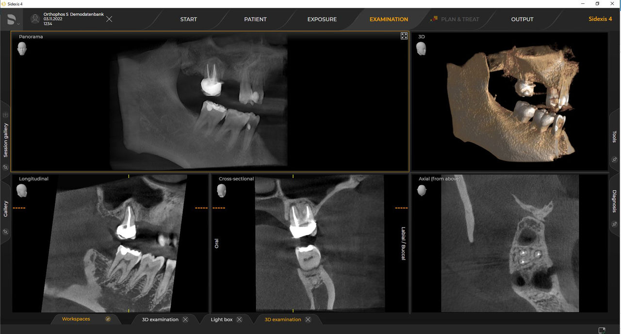 Ø 5 cm x 5.5 cm HD
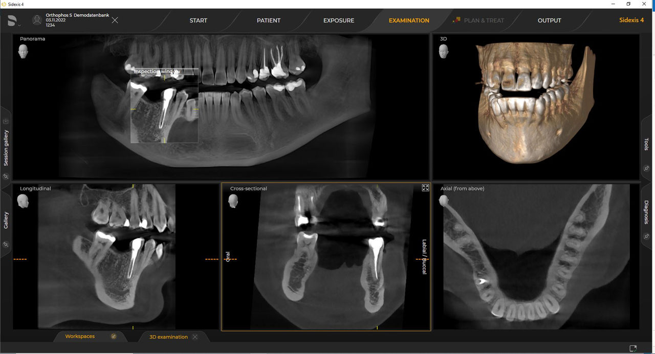 Ø 8 cm x 8 cm HD
 Ø 8 cm x 8 cm HD
Gallery of Cephalometric Images


FAQs
The requirements follow those for image processing software Sidexis 4 and Orthophos S. For more details please see Sidexis 4 system requirements and Orthophos S installation requirements.
Dentsply Sirona x-ray units work exclusively with Sidexis 4. Nevertheless data migration from Sidexis XG to Sidexis 4 is very easy. Sidexis 4 allows for the full digital experience with the latest tools
Orthophos S 2D allows a 3D upgrade. Axeos is a dedicated hybrid unit. The Orthophos E does not offer this option.


