


© 2025 - Dentsply Sirona, All Rights Reserved
The Intraoral Imaging Workflow
The Rinn intraoral imaging accessories from Dentsply Sirona, simplify the diagnostic workflow, helping clinicians work more efficiently while keeping patients comfortable and safe. They provide accurate positioning and control to consistently deliver diagnostically acceptable images in either digital or traditional media.
Diagnostic confidence makes a difference to the well-being of patients and the health of the dental practice. With the right tools and training, you can enhance the positioning and quality of diagnostic intraoral images while minimizing the risk. Read more below or contact us for more information.
Media + Aiming Devices - Autoclavable Solutions
Specialty Cases - Specialty Products
In some cases, maintaining patient comfort while capturing an accurate image requires an alternative approach. We offer holders to support all media types and sizes, for every case, setting clinicians up for success no matter what hurdles they may face.
Rinn Dental Aprons
The Soothe-Guard dental aprons are available in lead-free or lead-lined options with six premium colors and a magnetic closure option.
Benefits
Why choose Rinn intraoral imaging accessories?


Patient Safety and Comfort
Protect your patients and staff with Rinn products designed to prevent cross-contamination and minimize radiation exposure.
Autoclavable Solutions
For Digital Sensors and Phosphor Plate Holders, minimizing risks of errors and retakes to capture accurate images and increase patient comfort.
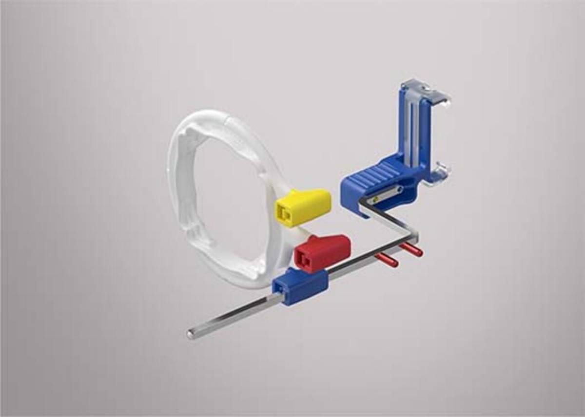
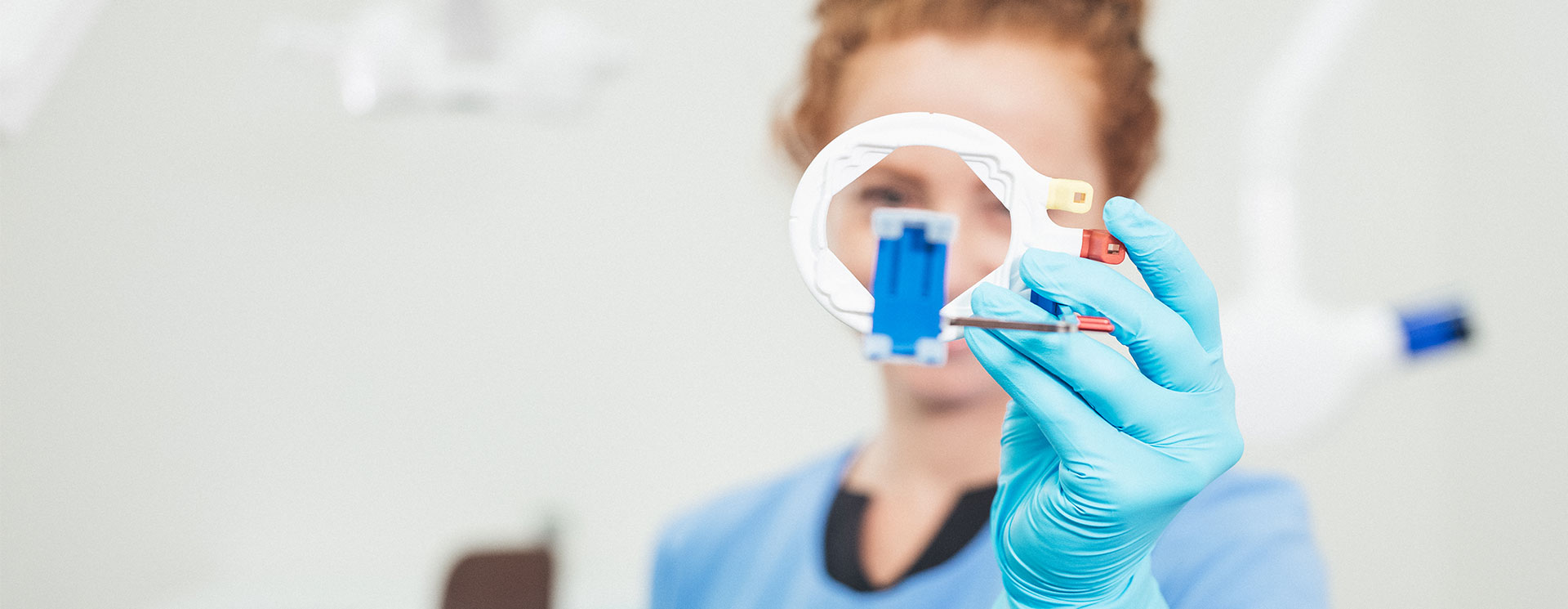
Color-coded positioning system
We offer products that work together with different choices of media to create a total solution.
Technology Equipment
Our imaging technology equipment simplifies the workflow for Digital Sensors and Phosphor Plate Holders and helps to capture diagnostically acceptable, high-quality images with improved efficiency.

ALARA Principal — As low as reasonably achievable
- Image quality and radiation exposure matter.
- The need for clear, diagnostically acceptable images should never supplant every clinician’s first principle: do no harm. That means keeping radiation exposure for your staff and patients as low as reasonably achievable: the ALARA principle. Best ALARA practices include:
- Utilizing rectangular collimation in conjunction with holders and aiming devices limits the central ray.
- Shielding patients with protective aprons and thyroid collars
- Minimizing retake images
- From radiation protection, to media holders and aiming devices, our products are designed to simplify your workflow, so you can focus on getting the image you need the first time
Tips and Education for Better Intraoral Imaging
The goal of dental radiology is to obtain high-quality images while minimizing radiation exposure and keeping every patient as comfortable as possible. Consider how these tips may assist in challenging situations.
- Know what to choose based on anatomical challenge.
- The paralleling technique using aiming devices is recommended for fewer errors and retakes.
- The bisecting angle technique using media holders are recommended for patients with anatomical challenges, and during endodontic and implant procedures.
- Use the largest sensor possible to obtain maximum information.
- Have a protocol in place for when to switch to a smaller sensor.
- Place in the center of the mouth for the paralleling technique.
- Use cotton rolls, especially in vertical positions and edentulous areas.
- Place the sensor parallel to the teeth.
- Use the indicator slot to align with interproximal spaces on bitewings.
- Align the central beam through the contact areas.
- Adjust vertical angulation on bitewings to capture adequate bone levels.
- With premolar bitewings, to capture the distal of the canine, adjust the media approximately 15 degress distomesial.
Downloads
Further downloads are provided within our Download Center.
- Acharya S, Pai K, Acharya S. Repeat film analysis and its implications for quality assurance in dental radiology: An institutional case study. Contemporary Clinical Dentistry. 2015;6(3):392-395.
