Specialty Cases
In some cases, maintaining patient comfort while capturing an accurate image requires an alternative approach. Anatomical challenges that benefit from special positioning techniques include:
- Shallow palates which may prevent the patient from occluding
- Narrow arches where an alternative media holder may be needed
- Patients with a sensitive gag reflex
Specialty Products
We offer holders to support all media types and sizes, for every case, setting clinicians up for success no matter what hurdles they may face.
For Digital Sensors
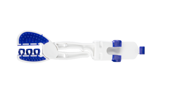
Rinn Snap-A-Ray DS Universal Sensor Holder
Easily capture anterior and posterior bisecting angle radiographs with one holder.
- Universal design fits all digital sensors
- Autoclavable for low cost per use
- Optional 2-in-1 arm and ring kit for alignment
- Rigid design for less movement provides accurate positioning
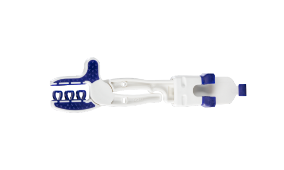
Rinn Snap-A-Ray DS Endo
Rinn Snap-A-Ray DS Endo for endodontic intraoral imaging has a cutout bite surface to leave room for your work.
For Phosphor Plates and Film
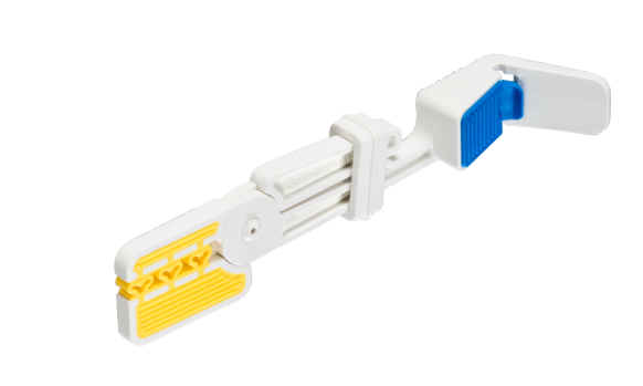
Rinn Snap-A-Ray Xtra Film & Phosphor Plate Holder
- Cushioned media grips hold film and phosphor plates securely without scratching the media
- Cushioned bite area designed to enhance patient comfort
- Media can be angled for hard-to-reach areas
- Easy to load and position media
- Autoclavable
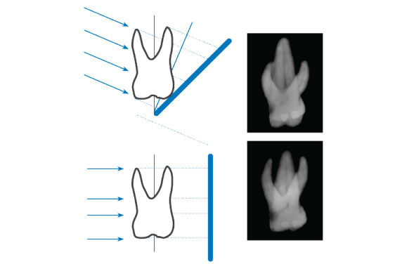
Tips: Bisecting Technique Steps
- Place the biteblock on the lingual aspect of the tooth
- Place the biteblock as close to the tooth being imaged
- The biteblock and long axis of the tooth form an angle
- Visualize an imaginary bisector line down the middle of the angle created by the tooth and biteblock
- Position the central ray perpendicular to the imaginary line
To see or download Instructions for Use, please visit our Dentsply Sirona IFU website and insert the product name in the search field.
Please contact us for more information about Preventive.