患者ポジショニングと撮影操作サポート
デンツプライシロナは患者ポジショニングと撮影操作を簡単にするための10ポイントコンセプトを開発しました。このコンセプトでは高品質な画像と患者さんの快適性の2点を主眼としています。このコンセプトにより、治療解析のための高品質な画像を確保するために必要なツールをサポート・提供し、患者さんとスタッフのための人間工学に基づく快適性を重視しています。
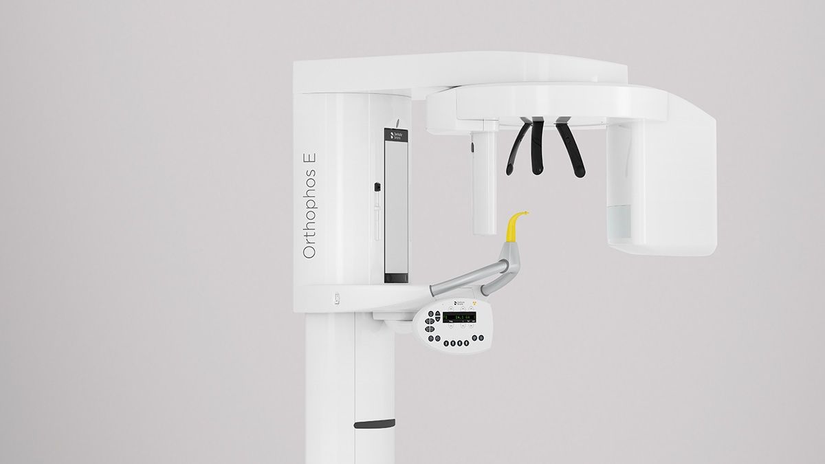
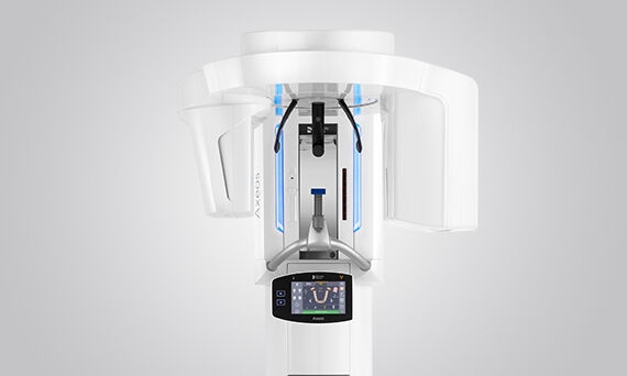
オーソフォスE 2Dは、CsIセンサーテクノロジーとその使いやすいインターフェイスにより、常に信頼性の高い診断を提供します。さらに、 セファロアアームのオプション選択することで、歯科矯正の信頼できるパートナーにもなります。 幅広いサービスであなたの診療を充実させてください。
一般歯科医および歯科矯正医向けのソリューションです。
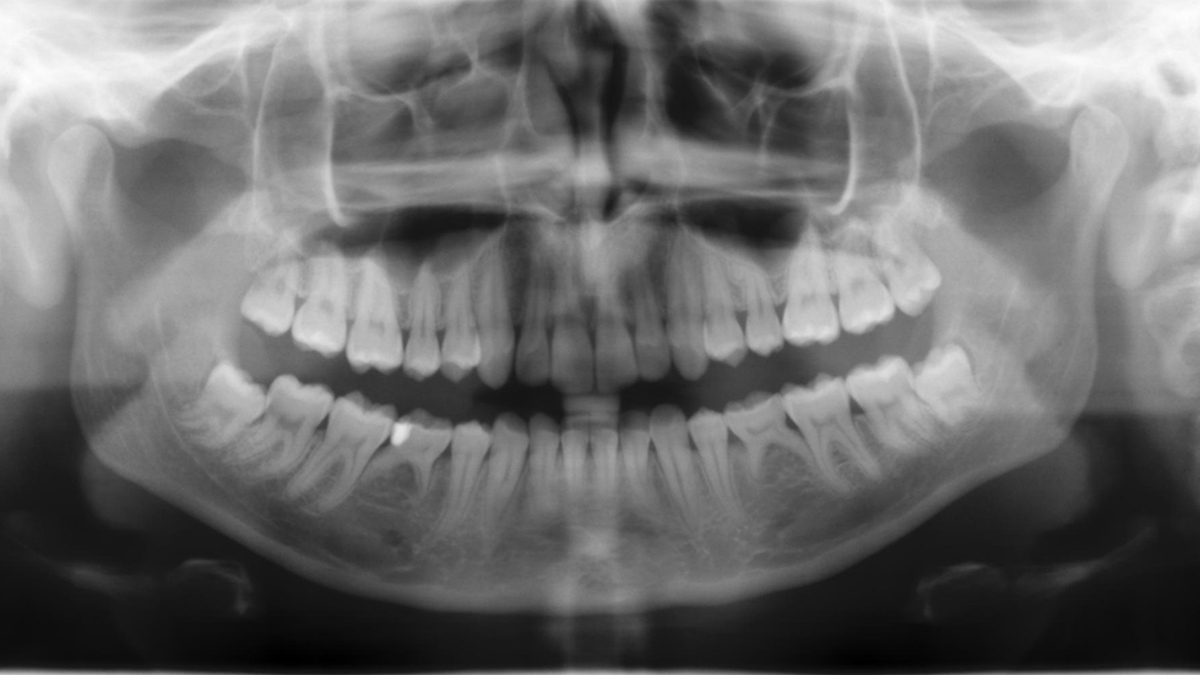
2D CsIセンサーで鮮明な画像が得られます。
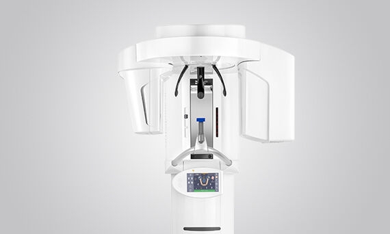
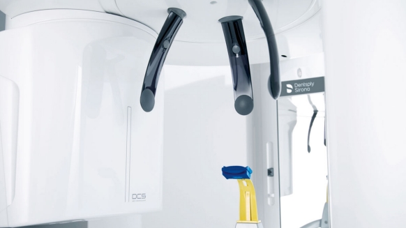
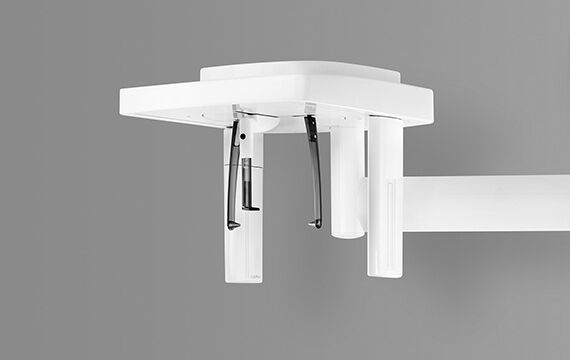
電動の3点式ヘッドサポートハンドルが患者さんをしっかり支え、同時にEVIビームライトが撮影範囲に含まれる患者さんの位置を示します。
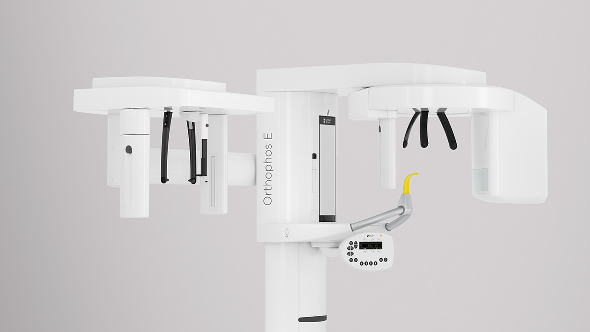
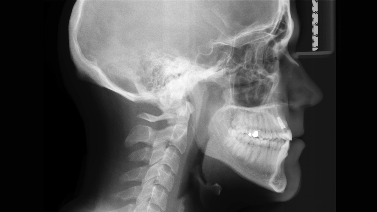
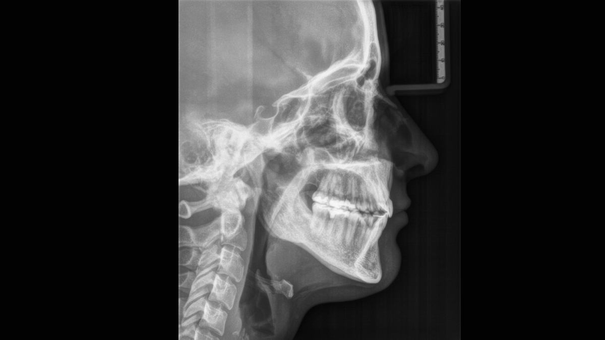
セファロアームは、オプションとして追加することも可能で、また、購入後いつでも後付けすることができます(レフトのみ)。
2Dの基本的な診断のためのプログラム。
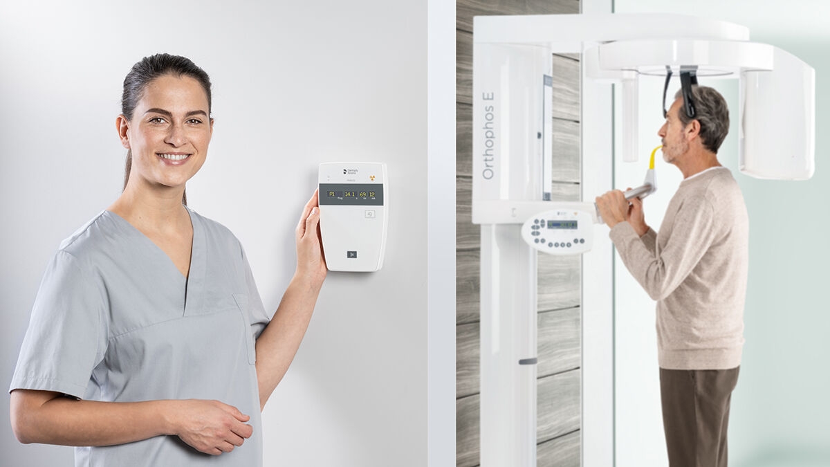
コントロールパネルで簡単操作が可能。

Given indications
Given indications
Given indications

Given indications


デンツプライシロナは患者ポジショニングと撮影操作を簡単にするための10ポイントコンセプトを開発しました。このコンセプトでは高品質な画像と患者さんの快適性の2点を主眼としています。このコンセプトにより、治療解析のための高品質な画像を確保するために必要なツールをサポート・提供し、患者さんとスタッフのための人間工学に基づく快適性を重視しています。
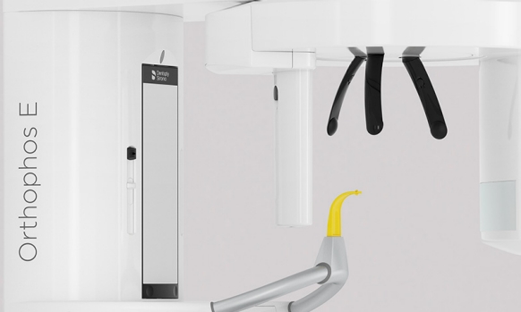
EVIビームライトが、患者さんの位置を示し、電動式の前額面とこめかみサポートが患者さんの頭部を固定。体動による画像のぼやけを防ぎます。
3D は、多くの歯科医院で認められつつありますが、多くの症例では依然、2Dイメージングが適切とされています。その大きな要因は放射線衛生学です。これは現代の歯科診断の中心となる構成要素です。画質の改善という観点では、2Dテクノロジーで利用可能なオプションはまだまだ尽きることはありません。デンツプライシロナ製の2D X線装置は、日々の取り扱いの面で高く評価されていますが、これは、驚くほど鮮明な画像と優れた操作性を可能とした技術的革新によるものです。
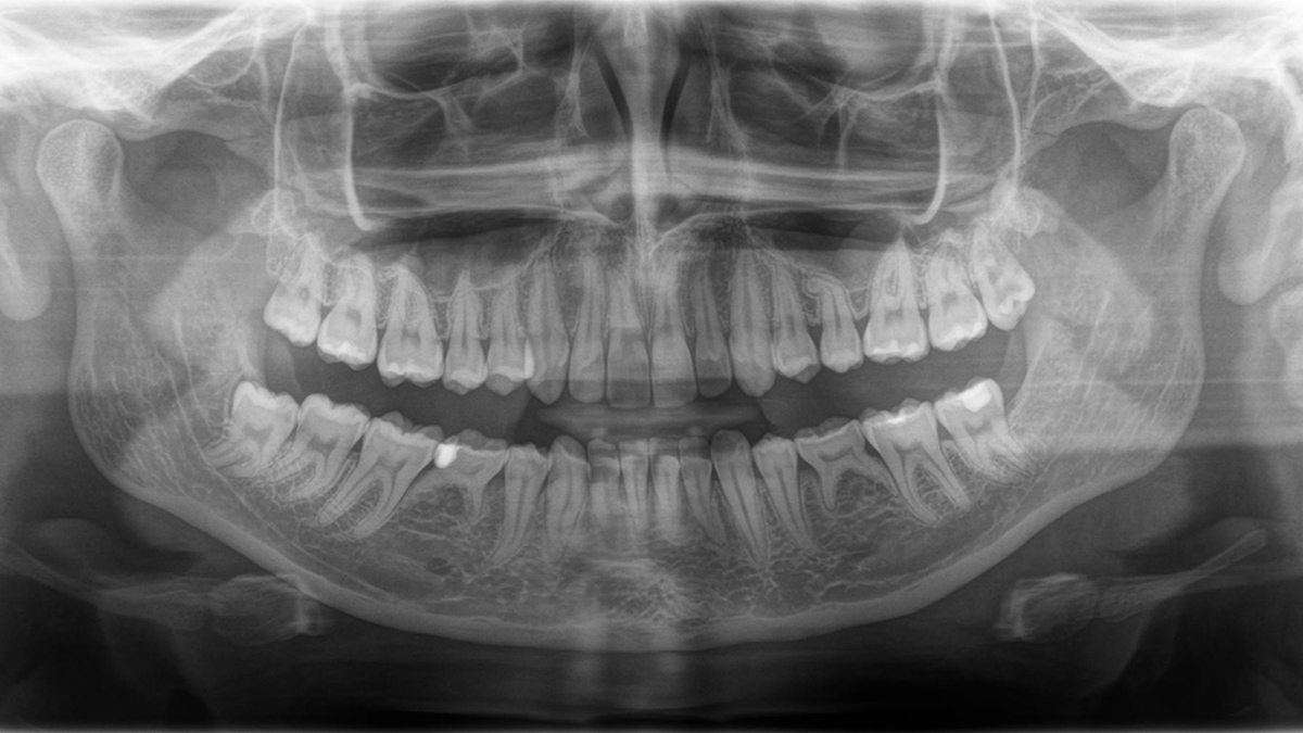
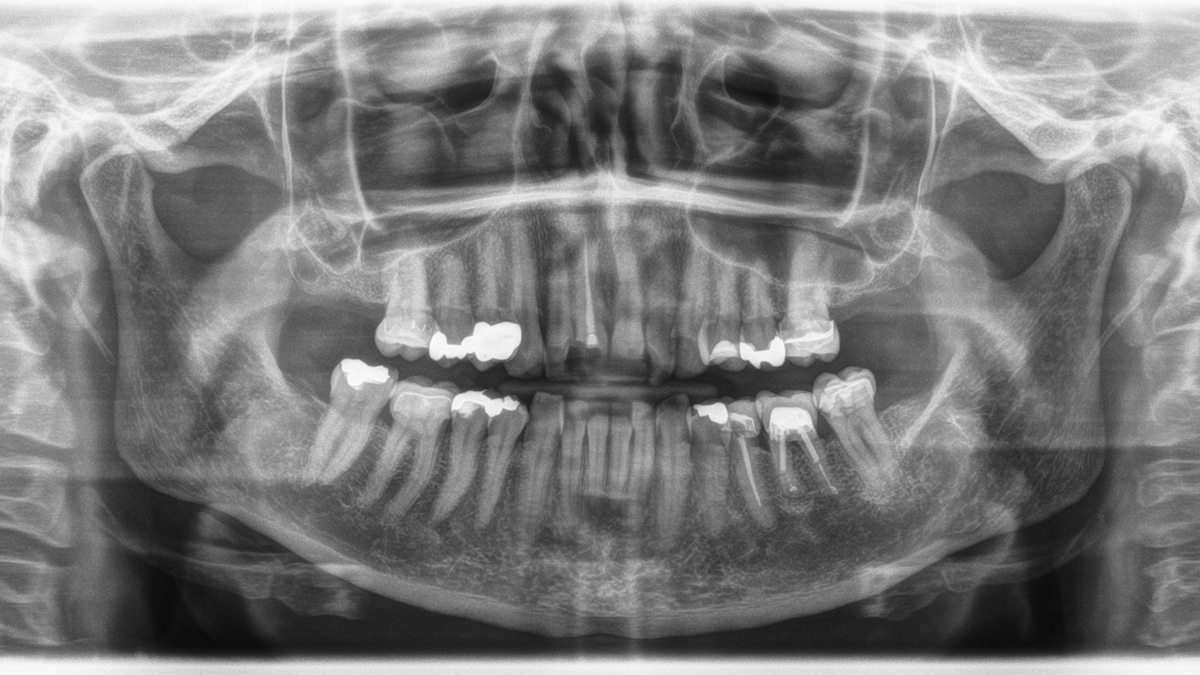
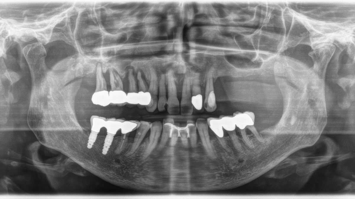
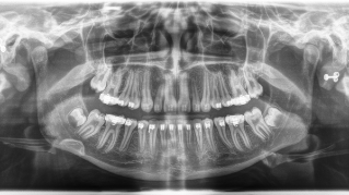
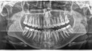
パノラマ画質は常に進化しています
左へ:10年が経過したPAN画像。右へ:オーソフォスSLで撮像したDCS画像
オーソフォスE 2Dには、いつでもセファロアームを取り付けることができます。(レフトのみ)


| Performance features | Orthophos S 2D | Orthophos E |
|---|---|---|
| X-ray generator | 60 - 90 kV, 3-16mA | 60 - 90 kV, 3-16mA |
| Panoramic exposure time | P1: max 14,2 s P1 Quickshot: max 9,1 s | P1: max 14,2 s |
| Radiation time Ceph | Standard 9,4 s Quickshot 4,7 s | Standard 9,4 s |
| User interface | EasyPad | MultiPad |
| Patient positioning | automatic (occlusal bite block) | manual |
| Panorama technology | CsI | CsI |
| Autofocus | yes | - |
| Ceph arm (optional) | left or right | left |
| Ceph unit with 2 sensors | yes | optional |
| Quickshot | yes | - |
| Fields of View | upgradeable | - |
| 3D Low Dose | upgradeable | - |
| HD mode | upgradeable | - |
| Base | optional | optional |
| Wheelchair accessible | yes | yes |
| Remote control | optional | optional |
| Ambient Light | - | - |
| Programme | Orthophos S 2D | Orthophos E |
|---|---|---|
| Standard panorama image | P1, P2, P10 | P1, P10 |
| Image detail left side or right side | P1, P1A, P1C P2, P2A, P2CP10, P10A, P10C BW1 | P1L, P1R |
| Image detail individual quadrants | P1, P1A, P1C P2, P2A, P2CP10, P10A, P10C | - |
| Image detail upper or lower jaw | P1, P1A, P1C P2, P2A, P2CP10, P10A, P10C, P12 | - |
| Constant magnification | P1C, P2C, P10C | P1C |
| Artifact-reduced | P1A, P2A, P10A | P1A |
| Thick layer front | P12 | P12 |
| Sinusoidal images | S1, S3 | S1 |
| Multislice in posterior tooth | - | MS1 |
| Mandibular joint | TM1.1, TM1.2, TM3 | TM1.1, TM1.2 |
| Bitewing image | BW1, BW2 | BW1 |
| Ceph (optional) | C1, C2, C3, C3F, C4 | C1, C2, C3, C3F, C4 |
はい、デンツプライシロナの3D X線装置はSIDEXIS 4でのみ作動します。しかし、SIDEXIS XGからSIDEXIS 4へのデータの移行は非常に容易です。
オーソフォス S 2Dは3Dアップグレード可能です。 オーソフォス Eはこのオプションを提供していません。