SICAT Implant guides you effectively and easily through the implantology workflow - whether you are an experienced implantologist or just starting out.
Seamless integration between software and hardware gives you a unique user experience and an effective partner for your workflow. SICAT Implant supports true precision and accuracy during the implant planning process. This helps the user anticipate possible complications and provides certainty and safety during the procedure.
In combination with the numerous surgical guide options, which can be ordered directly within the software, sent to your local laboratory or created directly on-site with your CEREC milling unit, you receive optimum support right up to the final implant restoration.
Advantages at a glance
SICAT Implant offers numerous advantages for your implant planning.
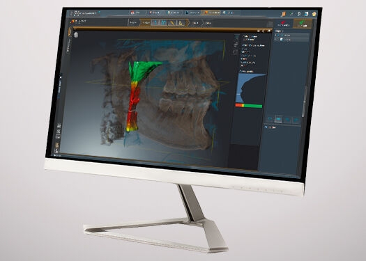
Clear and precise planning
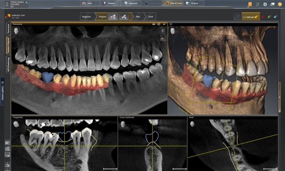
A clear user interface and intuitive planning tools give you everything you need for an efficient implant planning process. With the integration of your CEREC data, you can now view the entire planning process in realistic color and texture - for a truly unique planning experience.
Surgical safety and certainty
In combination with CEREC Guide 2 or 3, you can offer your patients high precision and a high degree of clinical safety. Whether you want to map the entire workflow in your practice, work with a local laboratory or use a third-party provider, SICAT Implant offers the flexibility to define the workflow that is right for you.
Higher treatment acceptance
3D visualization facilitates understanding and acceptance of the treatment plan for your patient and improves communication and the overall patient experience.
Guided processes are the future of implantology. They ensure greater precision and enhanced safety, especially in difficult situations.
Step by step: Implantology with SICAT implant
Pre-work: Scan
With a CEREC scanning system (Primescan or Omnicam), the gap situation (including antagonists) can be captured in minimal time. A prosthetic proposal for the gap situation is then planned. Thus, implant planning can be performed in a targeted manner, taking into account prosthetics, soft-tissue information and bone structure, for optimal accuracy and to achieve a functional and esthetic result.
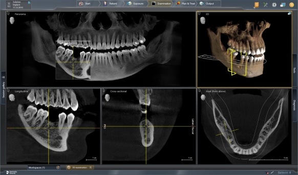
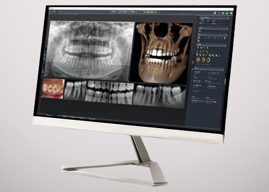
1. CBCT acquisition and diagnosis
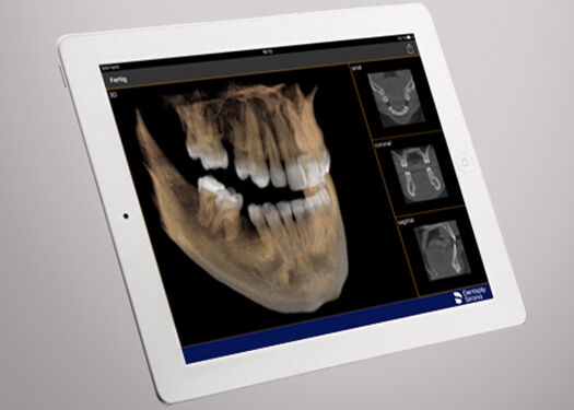
For a reliable diagnosis, it is important to be able to accurately examine the given situation from all perspectives. To get the right information, you first need the right equipment. Axeos, Orthophos SL 3D or Orthophos S 3D provide important 3D X-ray data needed to assess the clinical situation, and when partnered with the award-winning Sidexis 4 software your implant workflow is efficient from the first step.
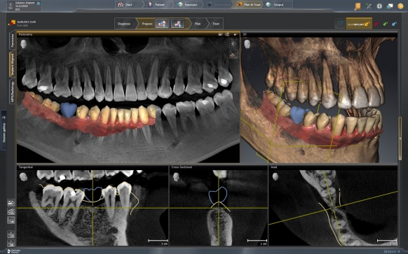
2. Superimpose the CAD/CAM data with the CBCT
If you are equipped with CEREC Omnicam or CEREC Primescan, you can import the digital impression and prosthetic proposals into SICAT Implant 2.0. The overlay ensures that the implant can be optimally planned according to the restoration axis.
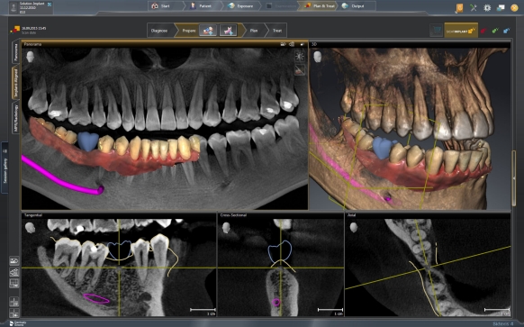
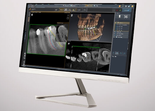
3. Map the nerve
SICAT Implant 2.0 offers you the ability to mark the nerve canal intuitively and with just a few clicks: Simply click on the nerve canal and continue along with individual clicks until you have successfully marked the entire nerve. Corrections (move, delete, expand, etc.) are uncomplicated and possible at any time.
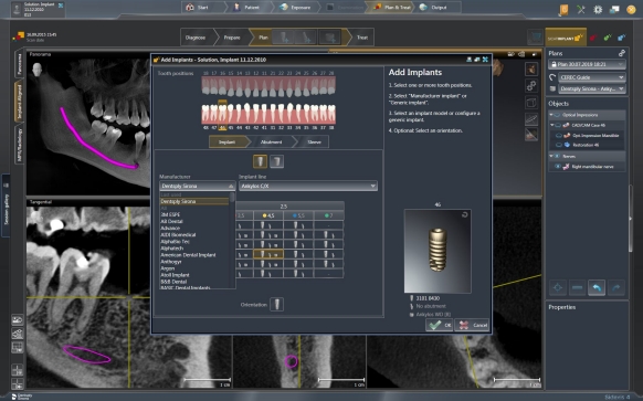
4. Select the implant
In addition to Dentsply Sirona implants (Ankylos, Astra Tech, Xive and MIS), SICAT Implant 2.0 maintains one of the most comprehensive databases of implants and abutments from major manufacturers. Regular updates are carried out in collaboration with numerous suppliers to keep the selection of implant systems up to date.
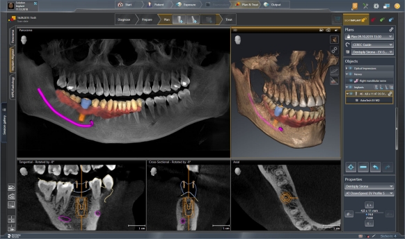
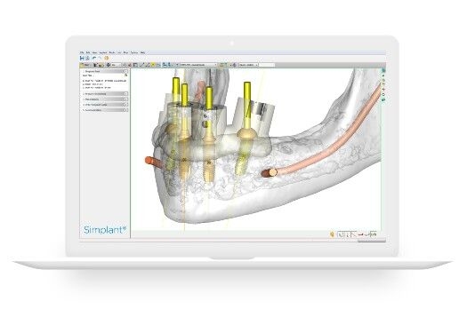
5. Position the implant
With just a few clicks, the implant is optimally positioned in the planning software in correlation to the bone. Thanks to the 360° rotation around the implant and the automatic collision warning in relation to the marked nerve canal and other planned implants, you can quickly achieve optimal placement. With the help of the prosthetic proposal, the implant axis can be optimized with the crown already in mind.
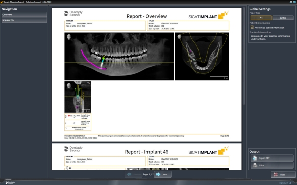
6. Write your report
With the available planning report, you can document the results of your implant planning with a single click and print them out for the upcoming surgery.
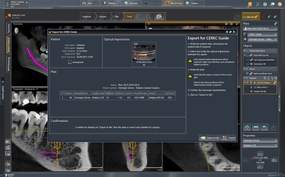
7. Surgical guide production
Depending on your needs, there are several ways to create a surgical guide to support the placement of the planned implant. You can order the template from SICAT directly in the SICAT Implant software, send it to a local laboratory or create a CEREC Guide with your CEREC Software and mill it directly with your CEREC milling unit in your practice. This allows you to remain flexible and decide which type of workflow you prefer.
SICAT Implant Tutorial Videos
Implant, Abutment & Sleeve Planning
Adjusting Panoramic Region & Measurements
Import & Register Optical Impressions
Marking & Adjusting Mandibular Nerves
Managing Plans
Imaging Solutions for Implantology
Learn more about Dentsply Sirona Imaging Solutions for Implantology