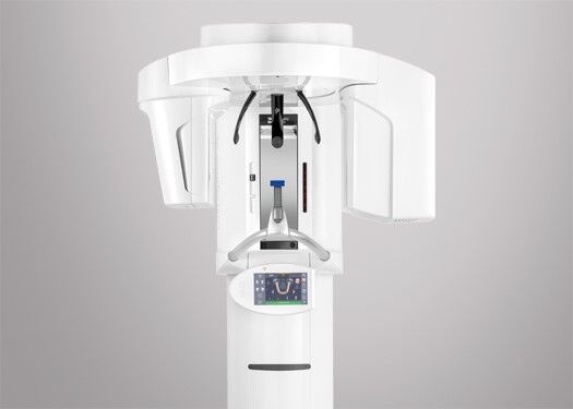
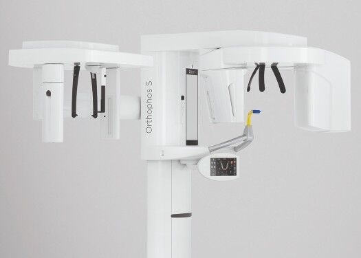
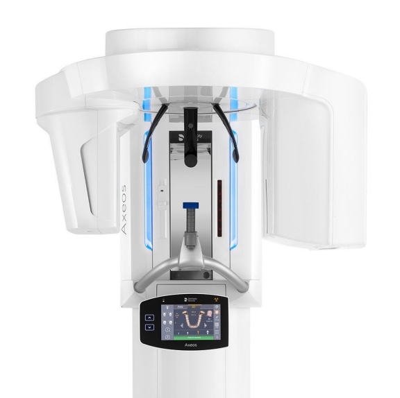
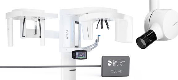
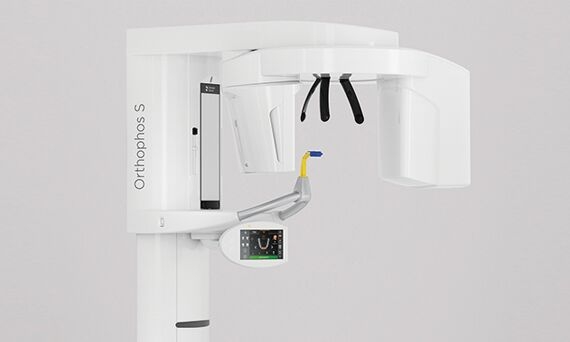
image-comparison grip-lines arrow-top-right zip arrow-right video minus arrow-back text shop twitter file home quick-order arrow-left youtube search arrow-down check all filter site_references image Slice 1 zoom 🎬 icons / _ icon size / _ download-queue Created with Sketch. chevron-down arrow-right picture pdf podcast exe plus hotline close xxx-1 burger clipboard linked-in ppt Artboard 🎬 icons / _ icon size / _ download-queue_add Created with Sketch. Layer 1 Created by potrace 1.15, written by Peter Selinger 2001-2017 My Account press_releases Created with Sketch. PRESS ds-instruments ds-preventive ds-implants ds-endodontics Cart presentation Created with Sketch. DS-Preventive-Icon-Grey video Created with Sketch. other Created with Sketch. text_piece Created with Sketch. promotion Created with Sketch. Spare Parts ds-imaging-systems case_study Created with Sketch. ds-treatment-centers broschure Created with Sketch. ds-prosthetics ds-restorative video Created with Sketch. flyer Created with Sketch. whitepaper Created with Sketch. article Created with Sketch. image Created with Sketch. Service ds-cad-cam Artboard Quick Order Dentist Chair study Created with Sketch. ds-orthodontics ds-implants ds-endodontics ds-cerec digital-scan grinder ds-diagnosis END-Website-Icons-Shaping-Cleaning-Obturation step_1 analyse ds-abutment-placement more-quality initiative scan more-quality ds-polishing digital ds-irrigation ds-treatment-centers Test treatment ds-restorative ds-implant-planning realistic construct book ds-planning-impression END-Website-Icons-Shaping-Cleaning-Obturation END-Website-Icons-Shaping-Cleaning-Obturation mirror anwender ds-preventive planning-2 ds-obturation ds-cad-cam 04 ds-implant-placement ds-retention-protection ds-anesthetics restauration form ds-inlab oven CORP-icon-grey-screen-tooth-cavity-pulpa robot plan place ds-orthodontics PDF-Formular_ganzneu PDF overlay ds-bone-regeneration ds-treatment-preparation vorher_nachher ds-maging-systems wifi END-Website-Icons-Shaping-Cleaning-Obturation omnicam PDF-Formular_neu_vonGünther treatment consulting planning comparison ds-set-up ds-prosthetics support step_3 ds-tooth-preparation ds-treatments ds-scaling step_2 ds-instruments software quote-english quote-french quote-text-german Created with Sketch. upload Created with Sketch. version Created with Sketch. ds-endodontics globe
Dentsply Sirona China
中文
主页 后退

Orthophos S


真正的全功能设备

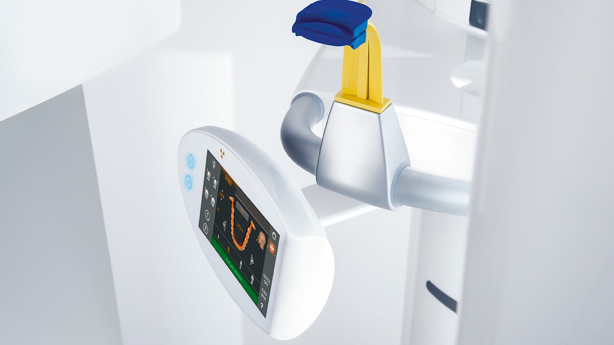
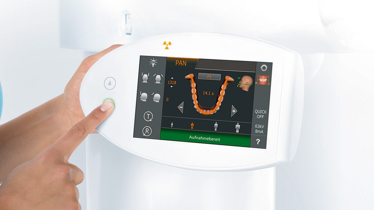
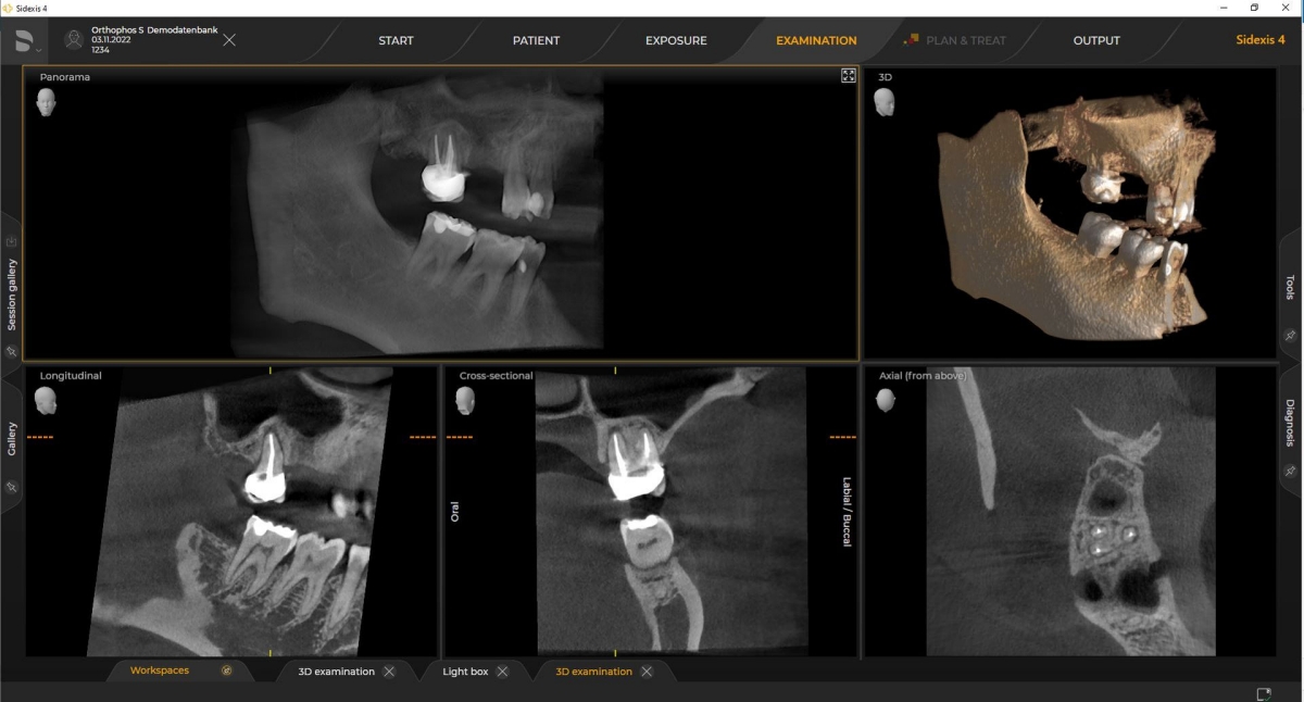
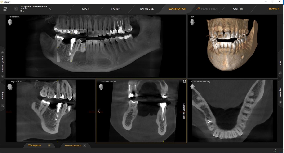
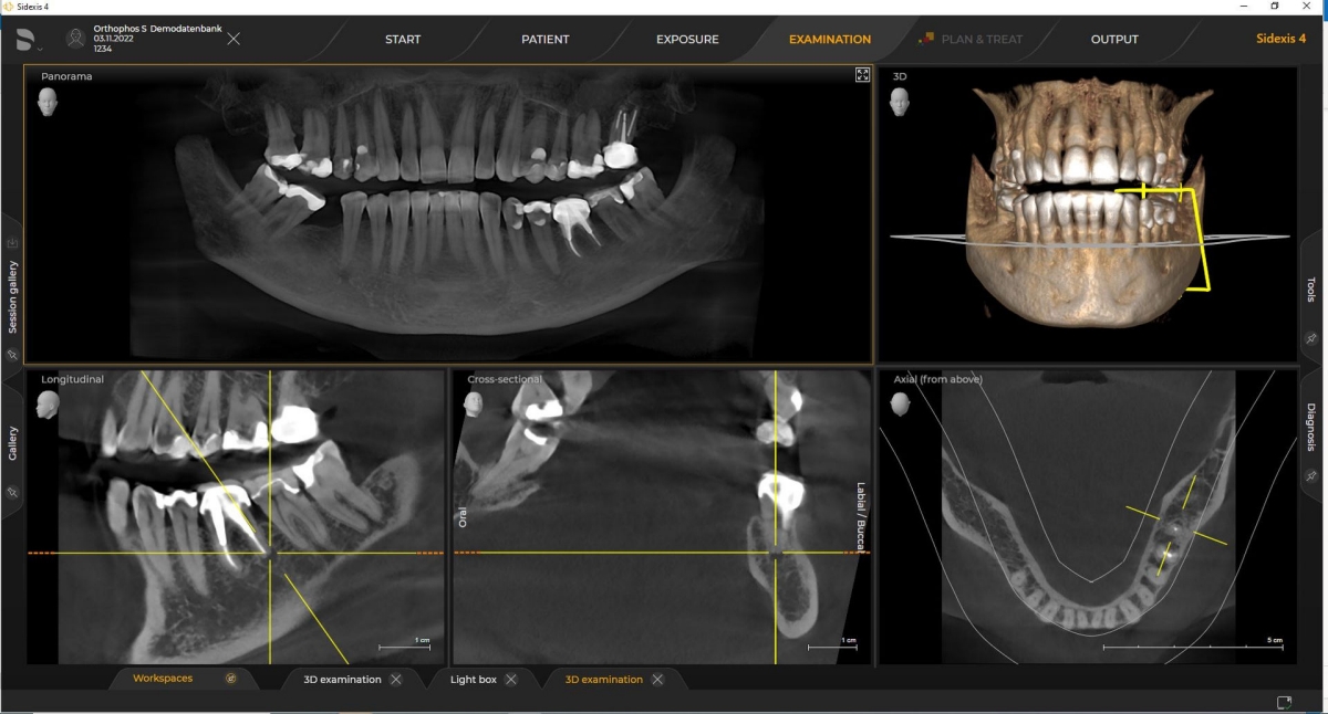
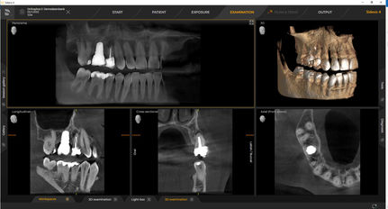
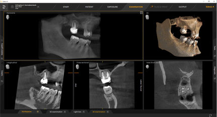
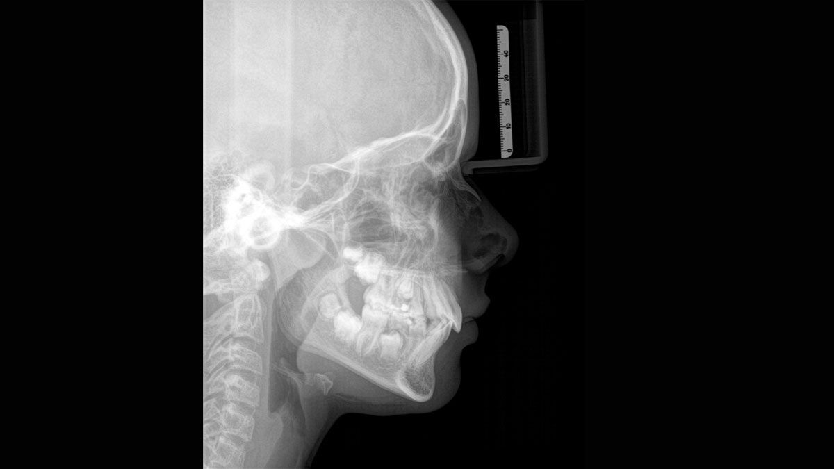
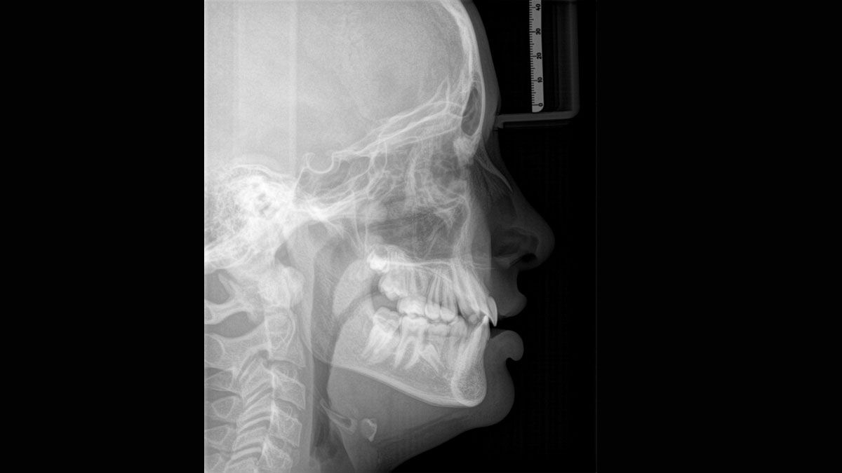
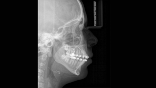
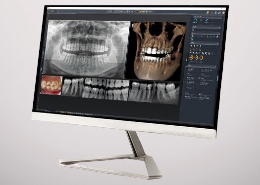
高质量的2D/3D X射线设备,能为每一家诊疗中心提供全面的服务。Orthophos S可以作为纯2D设备使用,也能提供3D建模,它具有可靠的性能,并且针对日常任务进行了优化。其Csl Plus传感器具有自动对焦功能,即使在颌关系复杂病例中也可确保图像清晰。通过借助已获专利的咬合块进行自动患者定位的功能,在很短的时间内就可以很方便地对患者进行定位。为了用于畸齿矫正,Orthophos S还提供了一个可选配的头影测量臂。登士伯西诺德始终坚持面向未来的发展战略,因此可以根据您诊所的需求随时加装头影测量臂。


登士柏西诺德的Orthophos S

Orthophos SL 3D, Orthophos S 3D

Orthophos S 2D

 
  • Panoramic exposure time: P1: max 14.2 s and P1 Quickshot: max 9.1 s
  • 全景照射时间:P1:最长14.2秒,P1快照:最长9.1
  • Ceph exposure time: Standard 9.4 s and Quickshot 4.7 s
  • 头影测量照射时间:标准:9.4秒,快照:4.7
  • User interface: EasyPad
  • 用户界面:EasyPad
  • Patient positioning: automatic (occlusal bite block)
  • 患者定位:自动(咬合块)
  • Panorama technology: Csl Plus
  • 全景技术:Csl Plus
Orthophos SL 3D, Orthophos S 3D

Orthophos S 2D/3D

升级到3D可获得这些附加功能:

  • Image volume (dxh)
  • 视野(dxh)

5cm x 5.5cm to 8cm x 8cm or 11cm x 10cm

5cm×5.5cm至8cm×8cm或11cm×10cm

  • MARS: Metal Artifacts Reduction Software
  • MARS:金属伪影消除软件
  • Intelligent Low Dose
  • 智能低剂量
  • HD mode

高清模式

Cephalometric Arm

Optional at the time of purchase with the Orthophos S 2D or 2D/3D hybrid unit, or it can be retroactively added at a later time.

购买Orthophos S 2D或2D/3D设备时的可选配置,也可以随时在以后添加。

The arm can be mounted on the right or left side of the unit based on your preference and space requirements.

该测量臂可以根据您的喜好和空间要求安装在设备的右侧或左侧。

Using a dedicated sensor, you get lateral and symmetrical p.a or a.p as well as carpus images

借助专用传感器,可以获得横向和对称的p.a.或a.p.以及腕骨图像。


为什么选择Orthophos S?

清晰且自动对焦的图像

2D Csl传感器具有自动对焦功能,即使在颌关系复杂病例中也可确保图像清晰

可选择的视野

直径5cm×5.5cm至8cm×8cm或11cm×10cm(可选)

可选的头影测量臂

头影测量臂可以添加在装置的右侧或左侧,可以在购买时订购,或随时加装

最大的一致性,最优的重复性

这都归功于借助已获专利的咬合块进行自动患者定位的功能

按需3D影像

在智能低剂量模式下,使用2D X射线相同的辐射剂量可获得CT的成像。在高清模式(高达1,400)下,在单次旋转过程中捕获单个图像,并将其转换为80μm的3D图像,以获得高分辨率的低噪影像

安全可靠的患者定位

自动太阳穴部位支架和前额支架,自动太阳穴宽度设置,激光瞄准装置和一体化手柄,可提高定位稳定性


易于使用。精确的患者定位。

对于大多数诊疗中心而言,使用X射线设备都是有两个主要目的:尽可能地捕捉到最佳图像,以进行准确、精确的诊断,以及确保患者在此过程中感到舒适。为了实现这两个目标,Orthophos S提供了独特且已获专利的解决方案:

  • The EasyPad and its intuitive user interface
  • EasyPad及其直观的用户界面
  • Automatic positioning aids such as the patented occlusal bite block and the 3-point head fixation
  • 自动定位辅助装置,例如已获专利的咬合块,以及3点头部固定装置
  • Integrated temple width measurement
  • 一体化太阳穴宽度测量

自动聚焦

正确的焦点对于拍摄优质的全景X光片至关重要。使用自动对焦功能,可自动得到具有最佳对焦清晰度的图像。登士柏西诺德成像设备可在一个周期内拍摄数千张图像,并自动识别下颌的最优定位的区域。这些图像随后会清晰地显示,而且无需任何手动操作。


不同视野选择

具有多种视野可选择(从直径5cm×5.5cm至直径11cm×10cm),可满足各种诊断和临床需求


Frequently Asked Questions

这些要求基于图像处理软件Sidexis 4和Orthophos S。有关更多详细信息,请参阅Sidexis 4系统要求和Orthophos S安装要求。

登士柏西诺德X射线机只能与Sidexis 4一起使用。不过,将Sidexis XG的数据迁移到Sidexis 4非常容易。Sidexis 4可使用最新的工具来提供完整的数字体验。

Orthophos S 2D可以进行3D升级。Axeos需要一个专用的升级配件。


联系我们

了解有关登士柏西诺德影像系统的更多信息,并索取有关口内图像、软件或 2D 和 3D 影像技术的产品信息。