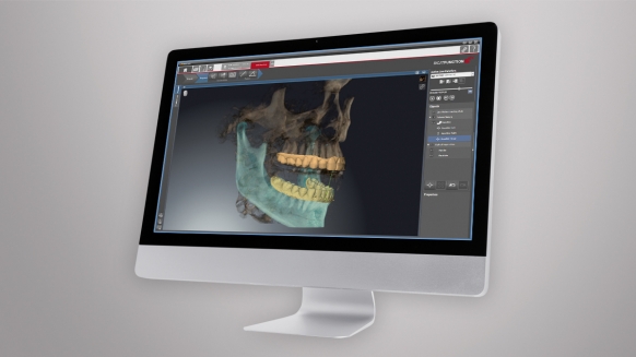
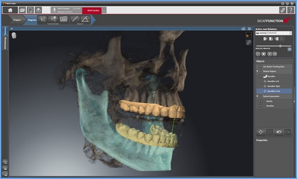
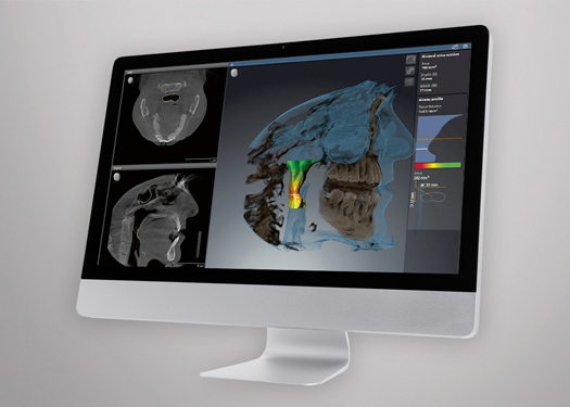
SICAT Function is the first integrated digital 3D solution to visualize real patient-individual movement of the lower jaw within the 3D volume. The anatomic traces of the temporomandibular joint can be displayed for every possible position in the volume.
Direct visualization of anatomically correct jaw movement, incl. real Condyle-Fossa relationship
Diagnosis, planning and consultation with your patient – all in one chairside session
Integration with CEREC for a 100% digital workflow
High patient acceptance
Your Benefits at a Glance
The new TMJD software for every practice.
Direct visualization of anatomically correct jaw movement
Real Condyle-Fossa relationship during jaw movement; Anatomically correct trajectory; Specific positioning of the trajectory in the 3D volume (if necessary also in comparison to conventionally used axial points); Evaluation of the occlusion based on the integrated optical surface scans
All in one chairside session
Diagnosis, planning and consultation with your patient.
Integration with CEREC for a fully digital workflow
Import your CEREC data for a purely digital workflow. You can also simulate the dynamic occlusion for each jaw position by means of the CEREC scans.
High patient acceptance
Integrated tools for patient education clarify the need for treatment.
SICAT Function in Action
TMJD Therapy with SICAT Function Step-by-Step:
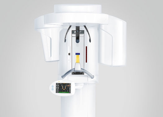
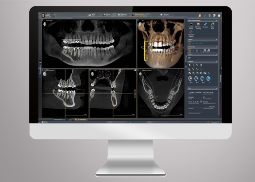
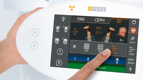
1. X-ray scan
The 3D X-ray data are recorded using Galileos Comfort Plus or Orthophos SL 3D unit.
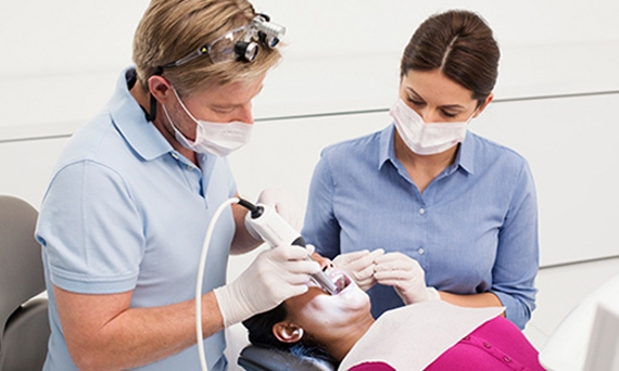
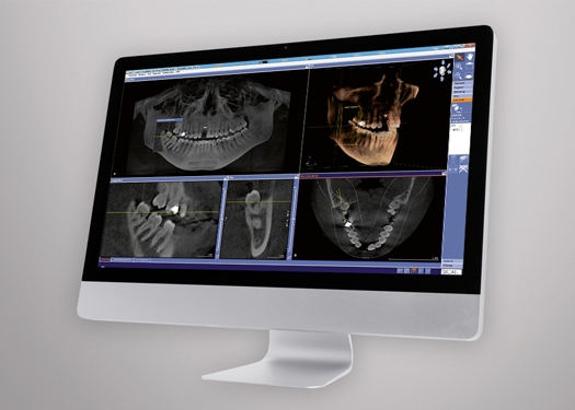
2. Record the optical surface
Recording the CEREC data of the patient's upper and lower jaw and subsequent fusion with the 3D data within the software.
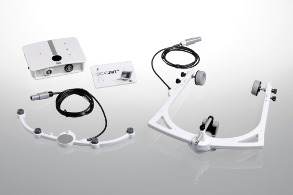
3. Record real jaw motion
Thanks to the highly precise recording of all degrees of freedom and movements of the mandible with the Jaw Motion Tracker SICAT JMT+, you can now transfer, visualize and diagnose anatomically correct jaw movement within the 3D volume:
Real Condyle-Fossa relationship during jaw movement
Anatomically correct trajectory
Specific positioning of the trajectory in the 3D volume – if necessary also in comparison to conventionally used axial points
You can also simulate the dynamic occlusion for each jaw position by means of the optical surface scans (CEREC).
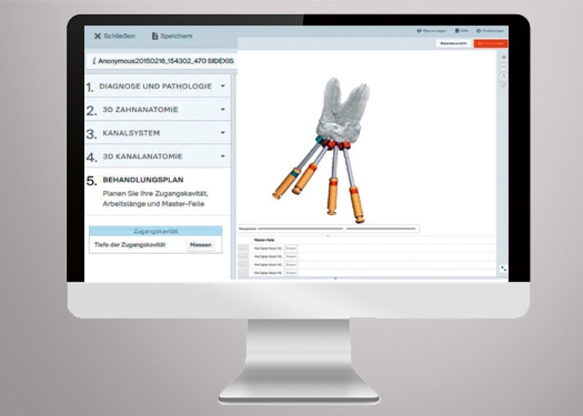
4. TMD diagnosis in four dimensions
Diagnostic information from the CBCT, the Jaw Motion Tracker (SICAT JMT+) and visual surface data (CEREC) are merged in SICAT Function. Thus, you get the first integrated digital 3D solution to visualize real patient-individual movement of the lower jaw within the 3D volume. The anatomic traces of the temporomandibular joint can be displayed for every possible position in the volume. In addition, the occlusion can be evaluated based on the integrated optical surface scans.
5. Order online the therapeutic appliance
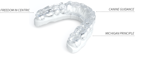
SICAT Optimotion is the first therapeutic appliance produced in a completely digital process using real, patient-specific jaw movement data.
The SICAT Optimotion therapeutic appliance is fabricated according to the principle of a Michigan appliance on the basis of the CBCT data, the optical surface scan and the jaw movement data from the SICAT JMT+. SICAT Function is the first solution to include both, the occlusion and mandibular joints in diagnosis and treatment planning.
Your benefits: customized therapeutic appliances that dramatically reduce the adjustment process of the appliance.
Dentsply Sirona CBCTs at a Glance
SICAT Function can be used with two models of Dentsply Sirona 3D X-ray units: Galileos Comfort Plus and Orthophos SL 3D.
Galileos Comfort Plus
The dedicated 3D X-ray unit, with a spherical 15cm volume and an optional Facescan for enhanced patient communication.
Dowiedz się więcej o systemach obrazowania Dentsply Sirona i poproś o informacje na temat obrazowania wewnątrzustnego, oprogramowania lub technologii obrazowania 2D i 3D.